| Home | Publications | People | Facilities | Links | Cytoskeleton J.Club |
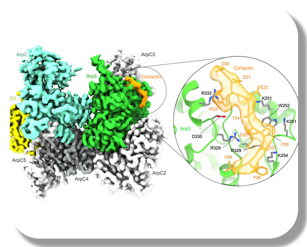
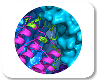
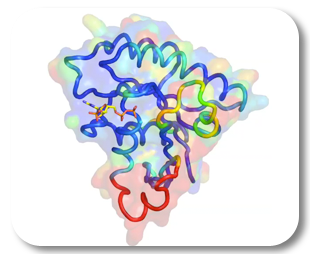
Cryo-EM structure of cortactin NTA-bound to Arp2/3 complex
Fregoso et al., 2023
PDB: 8TAH
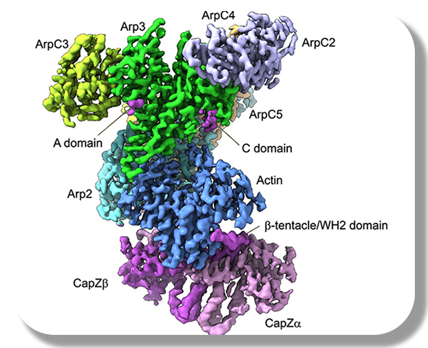
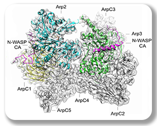
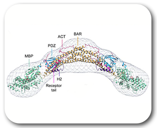
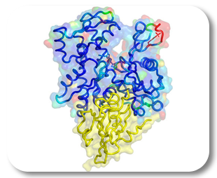
Transition State of Arp2/3 complex activation
VanEuwen et al., 2023
PDB: 7T5Q
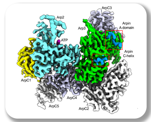
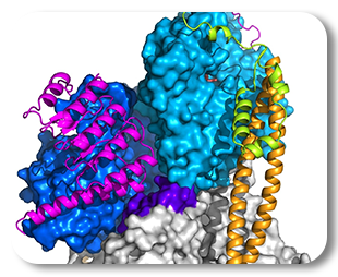
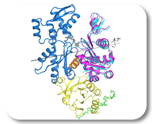
Carman et al., 2023
PDB: 8F8P, 8F8Q, 8F8R, 8F8S, 8F8T
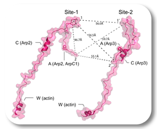
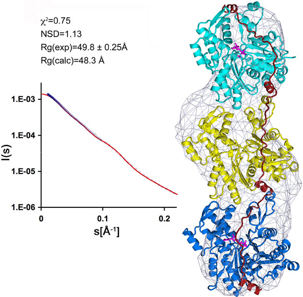
Fregoso et al., 2022
PDB: 7JPN
Lee et al., 2022
PDB: 6PSE, 6PSD
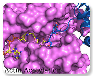


actin-profilin-Naa80
Rebowski et al., 2020
PDB: 6NBE, 6NAS, 6NBW
Zimmet et al, 2020
PDB: 6UHC
Kast et al., 2019
PDB: 6BCR
Lee et al., 2018
PDB: 6B9H
Turegun et al, 2018
PDB: 5TGC

capping vs. nucleation
Boczkowska et al., 2015
PDB: 4Z79, 4Z8G, 4Z94
Madasu at al., 2015
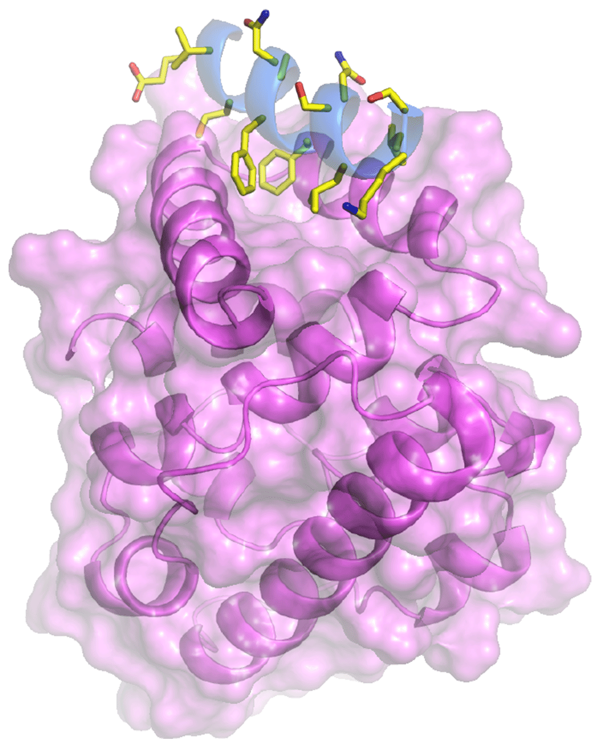
Rao et al., 2014
PDB: 4PKG, 4PKH, 4PKI
Kast et al., 2014
PDB: 4JS0
Boczkowska et al. 2014
Zwolak et al., 2013
PDB: 4K17
Madasu et al., 2013
PDB: 4J7O
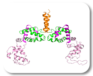
Rao et al., 2012
PDB: 3U1A, 3U1C, 3U5
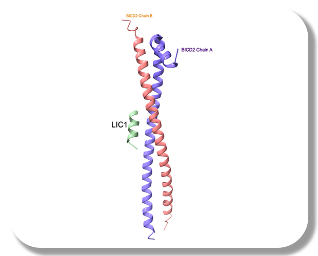
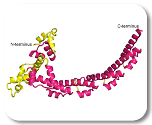
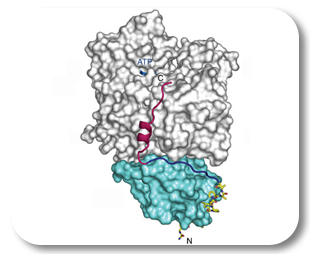
Namgoong et al., 2011
PDB: 3RYL
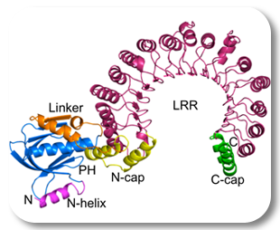
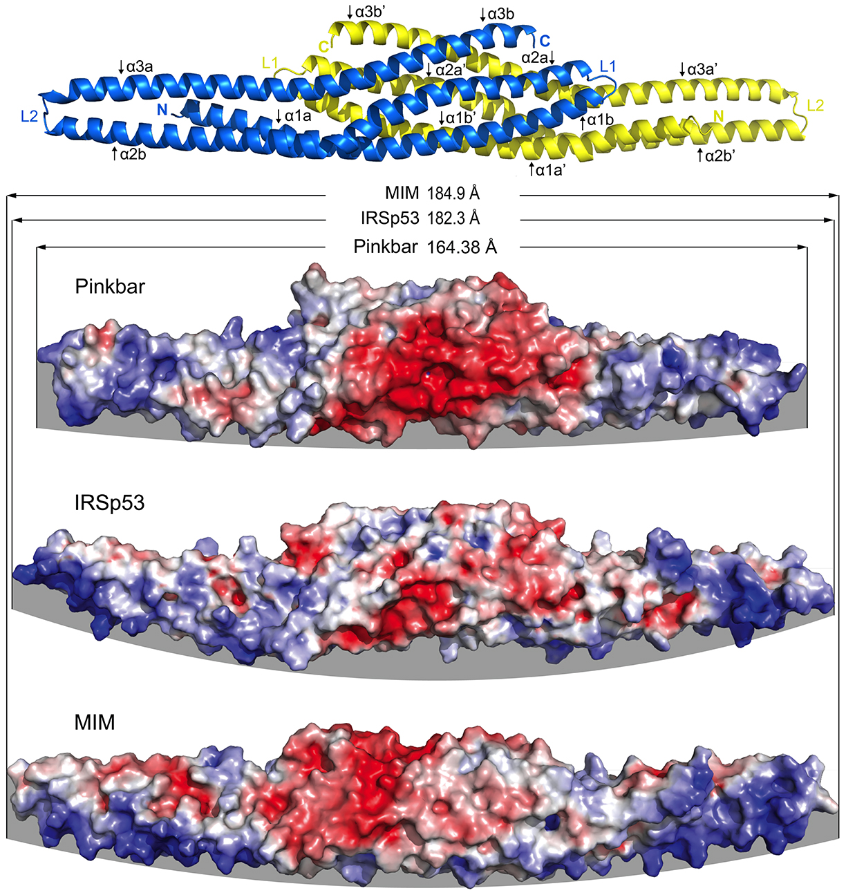
Pykalainen et al., 2011
PDB: 3OK8
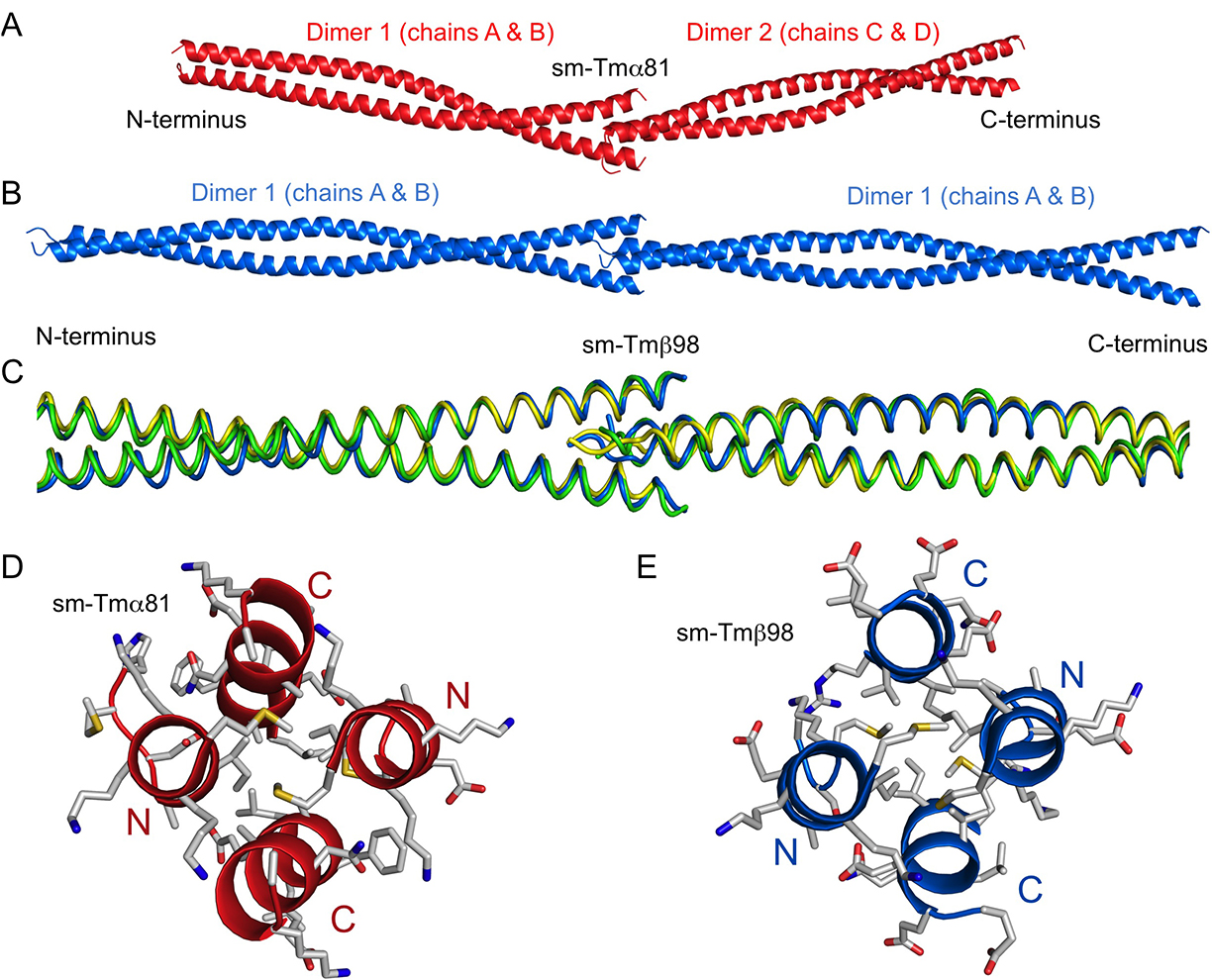
Jansen et al., 2011
PDB: 3P53
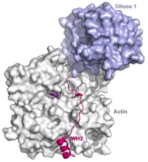
Rebowski et al., 2010
PDB: 3M3N
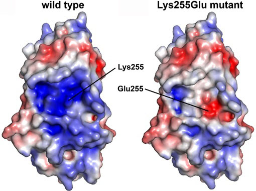
Baek et al., 2010
PDB: 3HIE
Lee et al., 2008
PDB: 3E5H, 2HXS
Baek et al., 2008

Baek et al., 2008
PDB: 3CHW
Boczkowska et al., 2008
Rebowski et al., 2008
Lee et al., 2008
PDB: 2R0O
Ferron et al., 2007
PDB: 2PAV, 2PBD


Lee et al., PNAS 2007
PDB: 2Q97
Lee et al., 2007
PDB: 2D1L
Chereau et al., 2005, Lee et al., 2007
PDB: 2A3Z, 2A40, 2A42, 2D1K
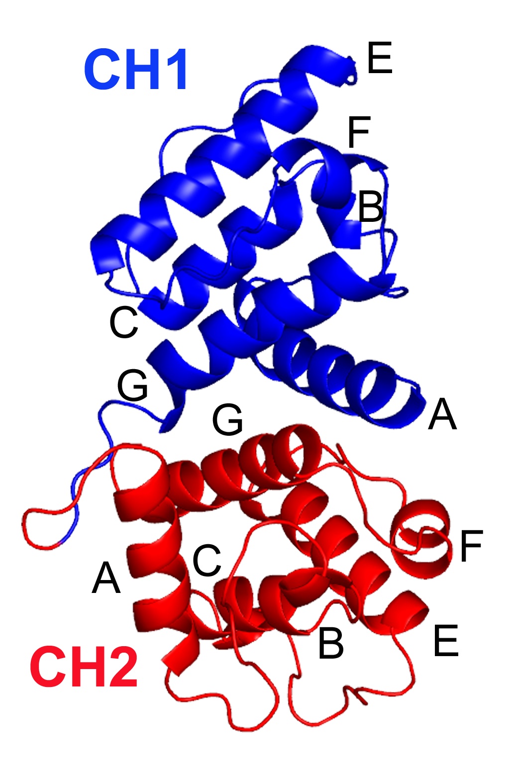
Borrego-Diaz et al., 2006
PDB : 2EYI, 2EYN
Terrak et al., 2005
PDB: 1N2D


(compact conformation)
Terrak et al., 2003
PDB: 1M46
PP1-MYPT1 complex
Terrak et al., 2004
PDB: 1S70
Otterbein et al., 2002
PDB: 1KXP
Otterbein et al., 2002
PDB: 1KW2
Otterbein et al., 2001
PDB : 1J6Z
Dominguez et al., 1998
PDB: 1BR1, 1BR2, 1BR4
Otterbein et al., 2002
PDB : 1K8U, 1K9P
Otterbein et al., 2002
PDB : 1K9K, 1K96