Research
Basic Research
In vitro cell lines are cultured and then implanted into rodents to create models for human brain tumors. Then, we can test various fluorophores on these models to identify those that could potentially help neurosurgeons in the operating room. This work allows us to study unique compounds and novel techniques in a pre-clinical setting (before patients) in preparation for clinical application. In addition, novel imaging technologies are tested in this setting in order to optimize visualization in humans.
Clinical Research
The goal of the clinical research is to perform intraoperative molecular imaging in an effort to optimize brain surgery.
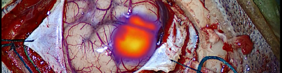
One technique relies on adaptation of an already FDA-approved dye that has been reformulated for tumor visualization -- Second Window Indocyanine Green (SWIG). This technique allows for real time intraoperative visualization of brain tumors and provides for visualization of tumor dye through normal brain parenchyma and intact dura. This nonspecific dye provides up to 1cm of depth penetration through normal tissues, including normal dura, thus allowing for early identification of tumors. The goal is for patients to benefit from the best possible tumor resection at the time of surgery.
For patients with pituitary adenomas, we have initiated a clinical trial using folic acid analog conjugated to a near infrared dye. Nonfunctioning pituitary adenomas overexpress folate receptor and thus provide an excellent target for this particular dye.
Future collaborations with both industry and nanoparticle bioengineering researchers include targeted, ph-sensitive, and nanoparticle dyes with theranostic capabilites as well.
Contact Us
Lee Visualization Lab
Department of Neurosurgery
Perelman School of Medicine at the University of Pennsylvania
235 South 8th St.
Philadelphia, PA 19107
215-829-6700
Email Us