The Perelman School of Medicine at the University of Pennsylvania
Office of the Executive Vice Dean and Chief Scientific Officer
2013 Art in Science Winners
We are delighted to announce the 2013 winners of our first annual “Art in Science” competition. We were pleased to receive a multitude of images from the Penn research community. We have awarded three prizes in each of the categories, Postdoctoral Fellows and Graduate Students. It is our pleasure to share the winning images below.
The full-sized images are currently on display in the EVD/CSO Suite.
The 2013 Perelman School of Medicine “Art in Science” Competition Winners
1st Place: Graduate Student Category
Amanda Phillips-Yzaguirre
Speck Lab
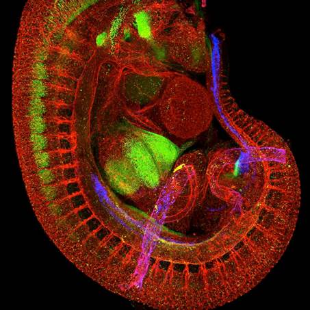
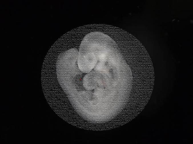
The image is a confocal Z-projection of the vasculature of an E11.5 transgenic (Ly6A-GFP) mouse embryo. Antibodies to 3 proteins were used in the production of this image. CD31 is shown in red it marks the vasculature of the embryo. Runx1 is shown in green it is expressed by blood cells and neurons in the somitic region. And GFP is shown in blue. Since this embryo is a Ly6A-GFP transgenic embryo the blue cells are those in which the Ly6A promoter is active. This image is a Z-projection acquired on a Zeiss LSM710 microscope. It is essentially an overlay of 96 images/channel so a total of 288 images which have been compiled to produce a 2D image of the entire vasculature of a mouse embryo (head and body wall of the embryo were removed prior to immunostaining).
The technique used to acquire this image is relatively new and requires the optical clearing of the sample using a benzyl benzoate/benzyl alcohol solution. I am interested in studying hemogenic endothelium. Hemogenic endothelium is a subset of endothelium that gives rise to all of the hematopoietic progenitors and hematopoietic stem cells (HSCs)during midgestation. Hemogenic endothelium expresses CD31, Runx1 and Ly6A. This image demonstrates that hemogenic endothelium is confined to the umbilical artery, the vitelline artery and the dorsal aorta. After hematopoitic cells arise from the hemogenic endothelium they migrate to the fetal liver where they undergo further maturation and proliferation. In the image you can see that the fetal liver is bright green with some blue spots, this is because hematopoitic progenitors retain Runx1 expression (green) and HSCs retain both Ly6A-GFP and Runx1 (blue and green) so from the image you can see that a majority of the hematopoietic cells in the fetal liver at this stage are not HSCs.
1st Place: Postdoctoral Fellow Category
Daniela Gómez Atria
Sunyer Lab
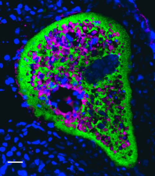
IgT mucosal immunoglobulin (green) is covering an Ichthyophtirius multifiliis trophont parasite (magenta) allocated in the skin epidermis of rainbow trout, 21 days after infection. Cell nucleuses are stained with DAPI (blue). Original magnification 200X. Scale bar, 20 μm.
This is the first demonstration of the existence of a specialized mucosal immunoglobulin in the skin of teleost fish.
2nd Place: Graduate Student Category
Efstathios (Stathis) D Gennatas, MBBS AICSM
Brain Behavior Lab
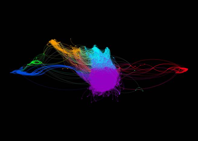
The figure consists of a force-directed layout of a brain graph based on the correlation matrix of 363 brain regions' gray matter density derived from 1074 subjects' T1-weighted MRI volumes (ages 9-23y.o.). This means that after looking at 1074 brains, regions whose gray matter density is highly correlated across subjects sit close together.
The graph is therefore a representation of part of the "common gray matter density covariance core" of the human brain. A modularity algorithm has been used to identify modules within the brain graph, i.e. groups of nodes with dense within-module connections and sparse between-module connections.
Node colors are based on module membership and edge colors are a mix between the colors of adjacent nodes. Modularity is a feature of complex networks like the brain and, at a glance, demonstrates that our node selection is meaningful and the defined networks are biological in nature and not artifactual.
Ongoing analyses are looking to establish normative sex-specific trajectories of development and correlations with neuropsychological test scores to explore the functional significance of these networks. Ultimately, aberrations in these systems will be related to brain disease and symptomatology. This is but a glimpse into our work to create sensitive and specific network-based neuroimaging biomarkers to further our understanding of normal and abnormal development and assist in the diagnosis and classification of brain disease.
2nd place: Postdoctoral Fellow Category
Benjamin Georgi
Bucan Lab
Essential genes are necessary for fundamental processes in an organism and lead to pre- or neonatal lethality when disrupted. In this work, we characterize 2,472 human orthologs of mouse essential genes in terms of their evolutionary and population genetics properties using data from recent deep sequencing initiatives in human populations. We find a signature of strong, purifying selection and reduced load of sequence variants within the putative essential genes when compared to a control-group of non-essential genes. We also show a significant enrichment of variants within essential genes across a set of four recent studies of de novo variants in patients with Autism Spectrum Disorder.
Our results establish the catalogue of putative essential genes as an important resource for analysis and interpretation of sequencing studies for human disease.
3rd place: Graduate Student Category
Kilangsungla Yanger
Stanger Lab
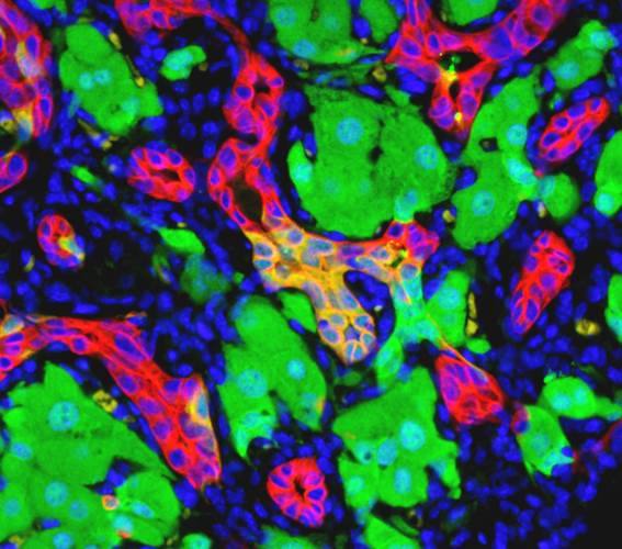
During its regenerative process, mature cells in the mammalian liver go through astonishing cell identity change known as ‘reprogramming.’ In this immunoflourescent image, hepatocytes in the mouse liver have been genetically marked with a yellow fluorescent protein (YFP, green) to lineage trace their eventual fate. When injured, they undergo morphological and molecular changes to express the marker, cytokeratin 19 (CK19, red), a marker for biliary cells (red) to reprogram into biliary cells and coalesce to pre-existing bile duct structures. This cellular reprogramming is a paradigm more closely associated with lower organisms until now, showing that the liver is capable of this in vivo under physiological conditions.
3rd place: Postdoctoral Fellow Category
Angela Conde
Weissman Lab
Human dendritic cells were nucleofected with HIV-1 envelope. Cells were then stained for HIV-1 envelope (purple), plasma membrane (red), and nuclei (blue) and analyzed by widefield microscopy.