Cardiac Imaging Group
-
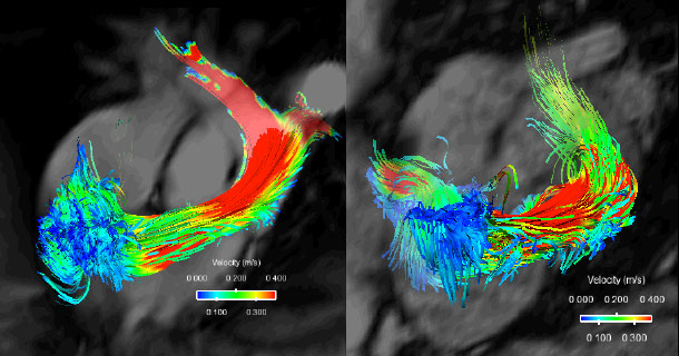
4D flow images in a normal subject (left) and a subject with pulmonary arterial hypertension (right).
-
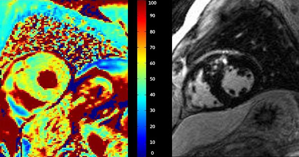
T1rho map (left) with high signal intensity in the same region of LGE (right) in a subject with hypertrophic cardiomyopathy.
-

Cine vs. real time imaging in two subjects (top and bottom) with arrhythmias. Graphs on the right depict the rhythm disturbance by examining real time volumes.
-
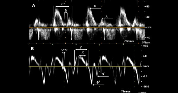
Assessing diastolic function using spectral Doppler and tissue Doppler.
Welcome
Our major interest is to use imaging tools such as echocardiography, cardiac MRI, and positron emission tomography (PET) to understand cardiac physiology/pathophysiology and to characterize the myocardium in different disease states.