Penn Center for Musculoskeletal Disorders
Uniaxial Testing
Uniaxial testing is a test in which a sample is subjected to a uniaxial force until failure. The uniaxial force can be applied as either a tension or a compression. During these tests, the forceapplied to the specimen is recorded as a function of displacement between the grips of the testing machine. Properties that can be directly measured via a uniaxial test are ultimate tensile/compressive strength, change in specimen length,and change in cross-sectional area.From these measurements the stress, strain, and Young’s Modulus (elasticity) of the specimen can be calculated.
The force measurement is used to calculate the stress using the following equation:

where F is the force and A is the cross-sectional area of the specimen.
The following equation is used with this to calculate strain:
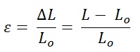
where delta L is the change in length of the specimen; L0 is the initial length of the specimen, and L is the final length.
The data points from these calculations can be graphed into a stress-strain curve, and the slope of the linear portion of this curve represents the Young’s Modulus (E) of the material. A typical stress-strain plot for a material that is tested to failure looks like this:
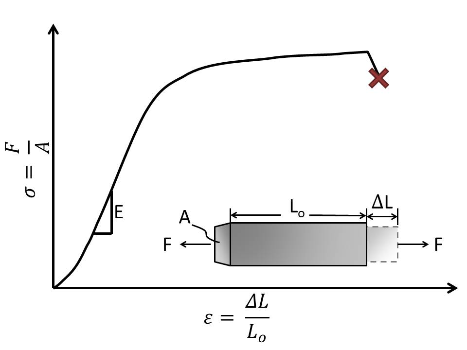
Figure 1: A typical stress-strain curve for a homogenous material that is tested to failure (X). The slope of the linear portion of the curve represents the Young’s Modulus (E).
Uniaxial Tension with Polarized Light:
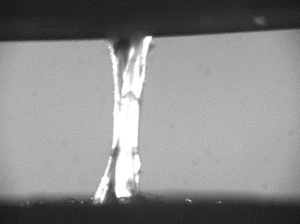
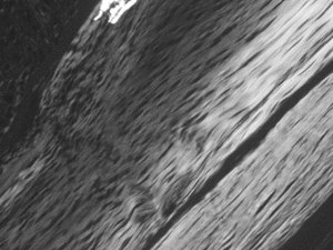
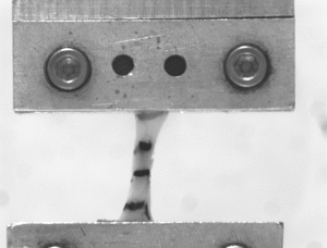
When examining the tendons of small animals, polarized light can be used during tensile testing to elucidate collagen fiber organization and alignment as a function of load. The birefringence of aligned collagen provides different amounts of illumination when exposed to varying degrees of polarized light. The intensity of illumination can be quantified for the entire tendon, which is correlated to the organization of the collagen within the tendon. Examples of tendons that are exposed to polarized light during tensile testing and under a microscope can be seen above.
© The Trustees of the University of Pennsylvania | Site best viewed in a supported browser. | Report Accessibility Issues and Get Help | Privacy Policy | Site Design: PMACS Web Team.