The Perelman School of Medicine at the University of Pennsylvania
Office of the Executive Vice Dean and Chief Scientific Officer
2015 Art in Science Winners
The Perelman School of Medicine Art In Science Competition
We are delighted to announce the 2015 winners of our annual “Art in Science” competition. We were pleased to receive a multitude of scientifically intriguing and aesthetically striking images from the Penn research community. We have awarded three prizes: one for Postdoctoral Fellows and two for Graduate Students. It is our pleasure to share the winning images below.
The 2015 Perelman School of Medicine “Art in Science” Competition Winners
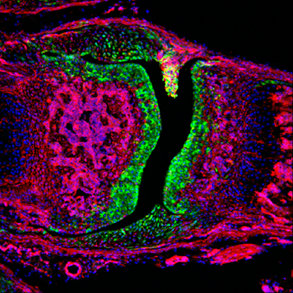
Winner: Postdoctoral Fellow Category
Rebekah Decker
Pacifici Lab
“Progenitor cells in the developing mouse synovial joint”
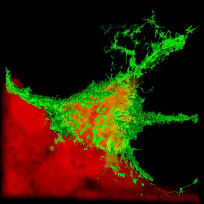
Jonathan Madara
Freedman Lab
Collaborators: Dr. Ron Harty and Dr. Gordon Ruthel
"Ebola Virus-like Particle Budding"
This is an image of green fluorescently tagged Ebola virus matrix protein (VP40) budding from an HEK 293T cell (the cytoplasm is counter-stained red). The matrix protein of many enveloped RNA viruses (including Ebola virus) is responsible for coordinating virion formation, and by expressing the VP40 matrix protein in culture, we can investigate mechanisms required for budding of Ebola virus.
Co-winner: Graduate Student Category
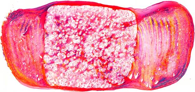
John Martin
Mauck Lab
“Stem-cell seeded engineered intervertebral disc in polarized light”
We harvested mesenchymal stem cells from cows and grew engineered intervertebral discs. This histology section is stained for collagen and is viewed under polarized to highlight the alternating layered structure in the outer region, mimicking the natural form of the intervertebral disc. Our finding was that stem cells were viable and continuously deposited extracellular matrix in longterm culture.