
Ultima Multi Photon Microscope
A multi-photon, or two-photon, microscope uses instrumentation similar to that of a laser scanning confocal microscope: A laser source for sample excitation, a scanhead with galvanometer controlled mirrors (or acousto-optic deflectors) to scan the excitation beam, and photomultiplier tubes to detect fluorescent signals. However, the confocal image differs in that optical sectioning is obtained using excitation in multi-photon microscopy whereas in confocal microscopy it is achieved using the emission pinhole.
The Prairie Technologies Ultima multiphoton microscope is similar in many respects to other available systems, but differs in that it contains two independent pathways of excitation, one for multiphoton imaging and the other for multiphoton uncaging. Because this system is additionally integrated with electrophysiology a diversity of applications are possible with our systems. For example, the fully configured system is able to perform 4-D imaging, to integrate imaging with photolysis, and to integrate 4-D imaging with photolysis and electrophysiology.
One of our Prairie Technologies Ultima systems attached to an Olympus BX-51 fixed stage microscope, is shown in the attached figure.
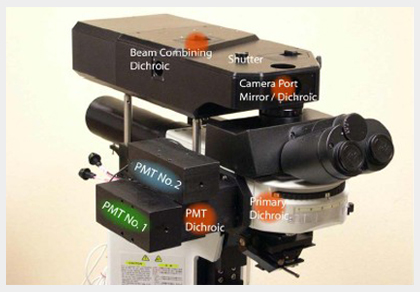
- Optical layout of Ultima Multi-Photon Microscopes
- What is the Two-Photon Process?
- Protocols
- Application Description
- Image Examples
- Instrumentation
- Sign-up For Equipment: Please contact Dr. Hajime Takano for further information.
Photo courtesy of Prairie Technologies, Inc.