
Pancreatic Ductal Adenocarcinoma (PDA) Models
Introduction
PDA development is accompanied by the formation of a unique tumor microenvironment, i.e., a dense and complex stroma that includes fibroblasts, blood vessels, and immune cells. It is well known that pancreatic cancer stroma is best modeled in autochthonous tumors, which arise spontaneously from the native locations as the result of genetic mutations. Therefore, to study stromal interventions, the genetically engineered mouse (GEM) models are strongly preferred.
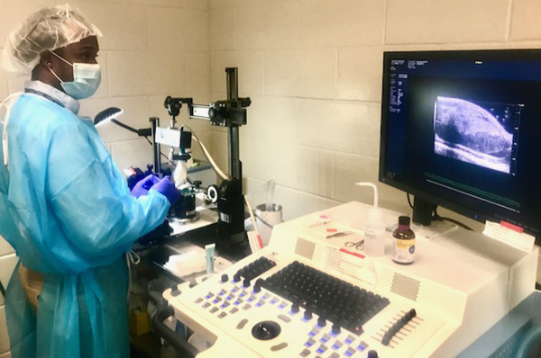
Mice harboring a pancreas specific Kras and p53 mutant with Cre alleles (KPC) was initially developed at Penn (Reference Paper) and is bred at the Mouse Hospital of Penn Pancreatic Cancer Research Center (For more information, download the Overview PDF).
Besides the GEM model, we have established orthotopic allograft model and xenograft (human PDA cell line) model. Both models are described in a Clinical Cancer Research paper, which compares the DCE-MRI results from KPC, orthotopic allograft and xenograft models.
Contact Us
Penn Quantitative Imaging Resource for Pancreatic Cancer
Perelman School of Medicine at the University of Pennsylvania
3620 Hamilton Walk
Philadelphia, PA 19104
Email: U24MRI@pennmedicine.upenn.edu
215-746-8747
Email Us
