Research
The gastrointestinal tract is constantly exposed to diverse infectious agents. Yet, despite the sustained presence of infectious agents in the gut, most individuals are not in a state of perpetual disease. Investigation of the trillions of bacteria that are part of the microbiota has revealed intricate ways in which our body endures the prolonged presence of these gut colonizers. In fact, we derive critical benefits from this symbiotic relationship. Bacteria help us break down nutrients and educate our immune system.
Compared with bacteria, however, little is known about how symbiosis is achieved with viruses and other types of infectious agents frequently found in the gastrointestinal tract. Our experimental findings indicate that mutually beneficial relationships between the mammalian host and diverse members of the gut microbial community are common, and that disrupting this balanced symbiotic relationship contributes to a range of chronic disorders. Below, we provide a brief description of several model systems we established in our laboratory, and how we are using them to learn about host-microbe symbiosis in the gastrointestinal tract and beyond.
1: Symbiotic viruses in the gut
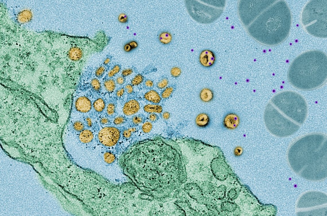
Noroviruses and many other intestinal viruses are routinely detected in healthy individuals. The consequences of these asymptomatic infections are unclear. We found that norovirus infection can be beneficial in mice by promoting the proper development of the immune system and protecting against intestinal injury, much like bacterial members of the gut microbiota. However, the same norovirus strain triggers intestinal abnormalities in mice that harbor a mutation in ATG16L1, a gene linked to Crohn’s disease. Crohn’s disease, along with ulcerative colitis, are the two major types of inflammatory bowel disease (IBD). These disease of the gastrointestinal tract are considered an abnormal immune reaction to gut microbes. They affect millions of individuals worldwide and remain difficult to treat. We are currently examining how norovirus and other viruses promote beneficial responses, whether this is mechanistically different from the way bacteria are beneficial, and why ATG16L1 mutation makes an otherwise innocuous virus become harmful. The answers to these questions will help us understand the origin of IBD and give us clues as to how to treat complex inflammatory diseases.
Live culture microscopy image of intestinal organoid “mini-guts” show that ATG16L1 protects against TNF-α, an inflammatory signaling molecule produced in response to norovirus infection. Control (left) and ATG16L1-deficient (right) murine intestinal organoids where grown in the presence of TNF-α and the cell death dye propidium iodide (red). Credit: Yu Matsuzawa-Ishimoto and Michael Cammer.
2: Autophagy and immunity
The Crohn’s disease susceptibility gene ATG16L1 is involved in autophagy, a process by which unwanted material inside cells are removed.We found that ATG16L1 and autophagy are important for protecting Paneth cells, a key cell type that protects intestinal stem cells, explaining why ATG16L1 mutant mice are susceptible to disease in response to viral infection. We and others in the field have discovered additional functions of ATG16L1 in immunity. Many of these functions involve autophagy, but not all. Together with the laboratory of Dr. Victor Torres at NYU, we recently discovered that ATG16L1 controls the release of small packages from cells called exosomes that act as decoys that soak up toxins made by bacteria such as methicillin resistant Staphylococcus aureus (MRSA). We are actively investigating how ATG16L1, autophagy, and autophagy-related processes protect against different disease-causing infectious agents.
3: Interactions between parasitic worms and the microbiota
According to some versions of the “Hygiene Hypothesis”, certain infections are important for the way our immune system develops. Eliminating these infections through improved hygiene may have increased our susceptibility to inflammatory diseases. In collaboration with the laboratory of Dr. P’ng Loke at the NIH, we found that infecting mice that harbor a mutation in NOD2, another gene linked to Crohn’s disease, with the parasitic worm Trichuris reverses disease by rearranging the bacterial microbiota. We also showed that Trichuris infection leads to similar rearrangement of the microbiota in indigenous people of Malaysia. These observations suggest that one way parasitic worms, also called helminths, promote the proper development of the immune system is by shaping the gut microbiota. We are in the process of characterizing helminth-associated bacteria to test this idea further. It is important to note that helminths infect billions of people in the world and can cause serious tropical diseases. For this reason, we are also interested in how the gut microbiota controls the helminth lifecycle, and whether we can target the microbiota for therapy against parasite infections.
4: Microbes acquired in the natural environment
Laboratory mice are kept in an ultra-hygienic environment and are not exposed to microbes the same way as humans in the real world. In collaboration with the laboratories of Dr. Andrea Graham (Princeton University) and Dr. P’ng Loke (NIH), we developed an innovative approach in which lab mice are released into an outdoor enclosure facility, a process that we call “rewilding” because we reintroduce mice into their natural habitat. We found that rewilded mice display a more mature immune system and changes to the microbiota, including a large increase in intestinal fungal colonization. We aim to use this setup to discover ways in which symbiotic microbes in the environment such as fungi drive immune development and disease susceptibility.