Perelman School of Medicine at the University of Pennsylvania
![]()
-
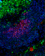
Confocal micrograph illustrating high levels of TACI on an extra-follicular foci (splenic sections stained with PNA, TACI, B220)
-
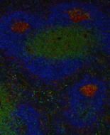
Confocal micrograph showing the absence of BLyS from the germinal center (splenic sections stained with PNA, BLyS, CD3ε)
-
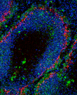
Confocal micrograph illustrating the anatomical location of germinal centers at the T-B border (PNA+ germinal center , CD3ε+ T, IgD+ B cells)
-
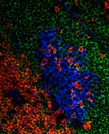
Confocal micrograph illustrating splenic architecture with MOMA-1+ macrophage layer seperating Marginal zone B cells (IgMhiIgDlo) from the Follicular B cells (IgMloIgDhi)
? Need help? Please feel free to e-mail us for more info.
Contact Us
284 John Morgan Building
3620 Hamilton Walk
Philadelphia, PA 19104
Office: (215) 898-8067
Fax: (215) 573-2350
Email: cancro@mail.med.upenn.edu
Faculty Web Page: Dr. Cancro