Clinical Areas
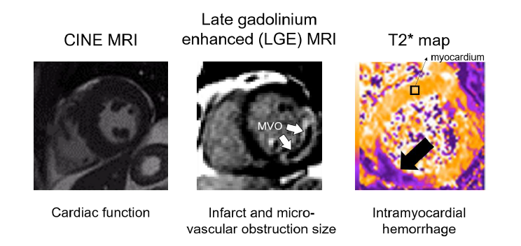
Ischemic heart disease is an enormous health and economic burden and the most common cause of death throughout the world. A devastating manifestation is acute myocardial infarction (MI) which results in myocardial loss and precipitates a cascade of events including myocardial scarring, adverse left ventricular (LV) remodeling, heart failure and death. Reperfusion injury is a frequent complication, and these patients have a greater risk of adverse tissue remodeling and cardiac function. While late gadolinium enhanced (LGE) MRI can detect myocardial fibrosis, there is significant interest in non-gadolinium contrast or endogenous contrast methods to spatially map infarcted tissue, detect recent ischemic injury and edema, or assess injury in patients with insufficient renal function who cannot receive contrast agents. Our team is developing non-invasive imaging and spectroscopic methods for myocardial infarction and reperfusion injury.
Tetralogy of Fallot
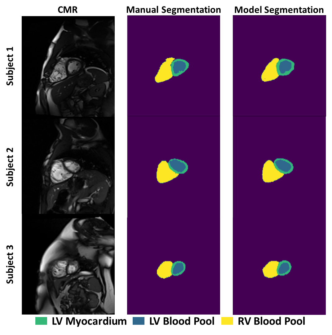
Tetralogy of Fallot (ToF) comprises 10% of congenital heart diseases and occurs 1 in 3600 births. After neonatal surgical repair, up to 44% of patients will go on to experience events related to pathologic right ventricular (RV) remodeling. These events include ventricular arrhythmias, RV dysfunction, and the need for re-intervention such as pulmonic valve repair. Despite significant advances in the overall survival of ToF patients, there is limited understanding of which patients will experience RV remodeling and adverse events. Our current projects focus on the applications of machine learning, computational fluid dynamics, and genetics to understand right ventricular remodeling in patients with ToF.
Collaborators: Mark Fogel, MD; Betsy Goldmuntz, MD
Single Ventricle Disease
Single ventricle disease (SVD) is an umbrella term for several CHDs which occur when a child is born with just one correctly-functioning cardiac ventricle. These patients often develop impaired cardiac function leading to significant morbidity and early mortality. Fibrosis is a major contributing factor to SVD patient outcomes, but its development is poorly-understood. We are working on multiple imaging-based methods to study SVD and its ramifications, including T1ρ mapping of the heart, as well as machine learning studies of function, fibrosis, and genetics.
Collaborators: Mark Fogel, MD; Betsy Goldmuntz, MD
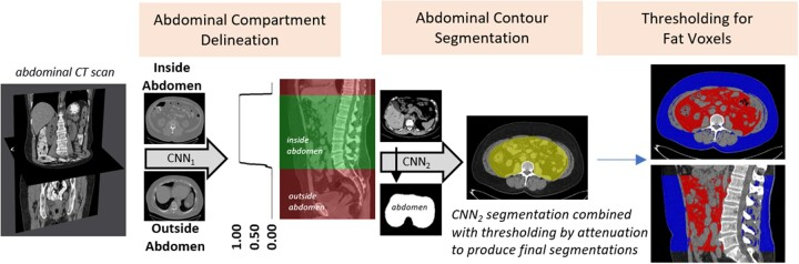
Non-alcoholic fatty liver disease (NAFLD) is a pathology of epidemic proportions. It affects approximately one-third of US adults and is a major cause of cardiovascular disease-, cancer- and cirrhosis-related mortality. While some genetic variants are known, they explain only a small fraction of its high heritability and many variants remain undiscovered. Widespread strategies to identify patients with NAFLD are needed, especially with early subclinical disease, and to conduct research studies of its genetic determinants. Our long-term goals are to develop easily accessible, non-invasive imaging strategies to identify patients before they progress to advanced liver disease.
Collaborators: Dan Rader, MD; Hersh Sagreiya, MD
Friedreich's ataxia (FA) is a debilitating disease caused by a deficiency in mitochondrial protein frataxin. Frataxin loss has a deleterious effect on cellular energetics, manifesting in multiple pathologies including cardiomyopathy. NAD+ supplementation may improve mitochondrial energetics and is being actively investigated. Our group is collaborating with researchers at the Children’s Hospital of Philadelphia to investigate MRS techniques to measure NAD+ at baseline and following NAD+ supplementation to show that NAD+ augmentation in brain, skeletal and cardiac muscle is associated with an improvement in function.
Collaborators: Shana McCormack, MD; Kimberly Lin, MD
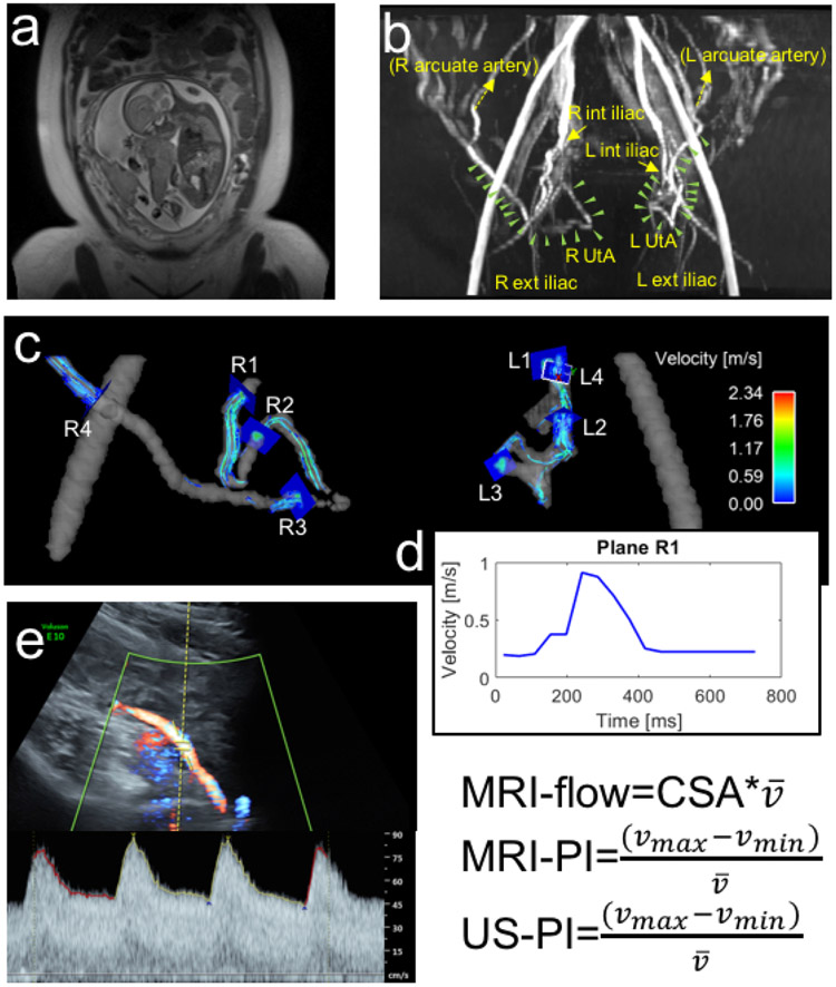
The National Institute of Child Health and Development recently launched the Human Placenta Project, a nationwide collaborative research effort to study the placenta and its effects on both the mother and the fetus. At the University of Pennsylvania and Children's Hospital of Philadelphia, the Examining Placenta Oxygenation Perfusion and Size (EXPLORE) team (PIs Nadav Schwartz and Daniel Licht) consists of clinicians and imaging scientists working to assess how in vivo development of the placenta can impact management of maternal nutrition. Our role is to develop flow and perfusion imaging techniques of the placenta using phase contrast (i.e. 4D flow) and arterial spin labeling MRI approaches.