MRI Technology Development
Proton magnetic resonance spectroscopy (1H MRS) provides noninvasive, quantification of in vivo metabolism in healthy and diseased tissues. While conventional “upfield” MRS (0 to 4.7 ppm) is well-established and shows several key metabolites, there is much less established methodology regarding detection of “downfield” MRS (>4.7 ppm) metabolites such as nicotinamide adenine dinucleotide (NAD+), phosphocreatine (PCr), adenosine triphosphate (ATP), glutamine (Gln) and carnosine in vivo. We are actively developing MRS pulse sequences and post-processing strategies at 7 and 3 T in order to detect and characterize downfield metabolites.

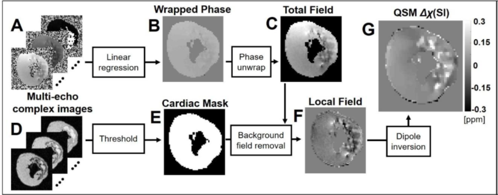
Quantitative Susceptibility Mapping (QSM) is a new MRI technique that permits noninvasive assessment of the magnetic susceptibility of tissues and can target tissue iron. MRI imaging of myocardial magnetic susceptibility can show abnormal metabolism of iron in cardiovascular disease, aging, or during development. QSM is a challenging mathematical inverse problem. MRI can measure the induced magnetization in tissues, which is proportional to applied magnetic field. By measuring the induced magnetization, we can uncover the tissue magnetic susceptibility by solving the inverse problem.
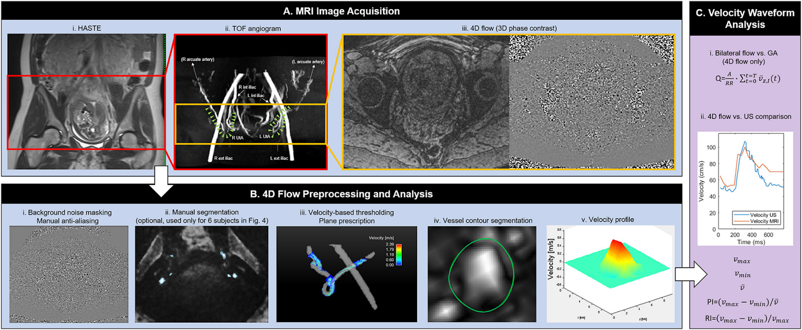
Phase contrast imaging is a type of MRI which enables measurement of velocity via changes in phase. 4D flow imaging is a type of phase contrast imaging, performed with velocity encoding in three spatial directions, and over time (the 4th dimension in “4D”). Consequently, 4D flow MRI is a valuable tool for assessing cardiovascular physiology through measurement of blood flow throughout the body. Computational Fluid Dynamics (CFD) is a complementary set of tools by which flow can be calculated and analyzed. Our group has previously used 4D flow combined with CFD to assess flow in the uterine arteries to study hemodynamic factors in the pathophysiology of preeclampsia.
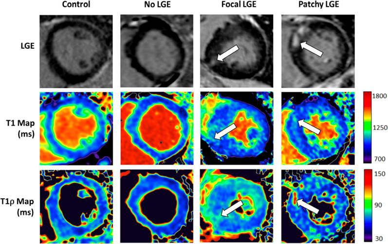
Our group is investigating T1ρ (“T-one-rho”) MRI as an endogenous contrast method for quantitative non-invasive imaging of myocardial injury. In comparison to T2 and native T1, T1ρ permits better detectability of myocardial fibrosis with improved specificity. Our research is investigating the origins of endogenous contrast in myocardium, fibrosis detection in large animal models of ischemia reperfusion and in patient studies. Our recent work developed new methods for imaging infiltrative fibrosis in hypertrophic cardiomyopathy and early studies demonstrating the potential for detection of interstitial disease.