Artery Development
Artery Development
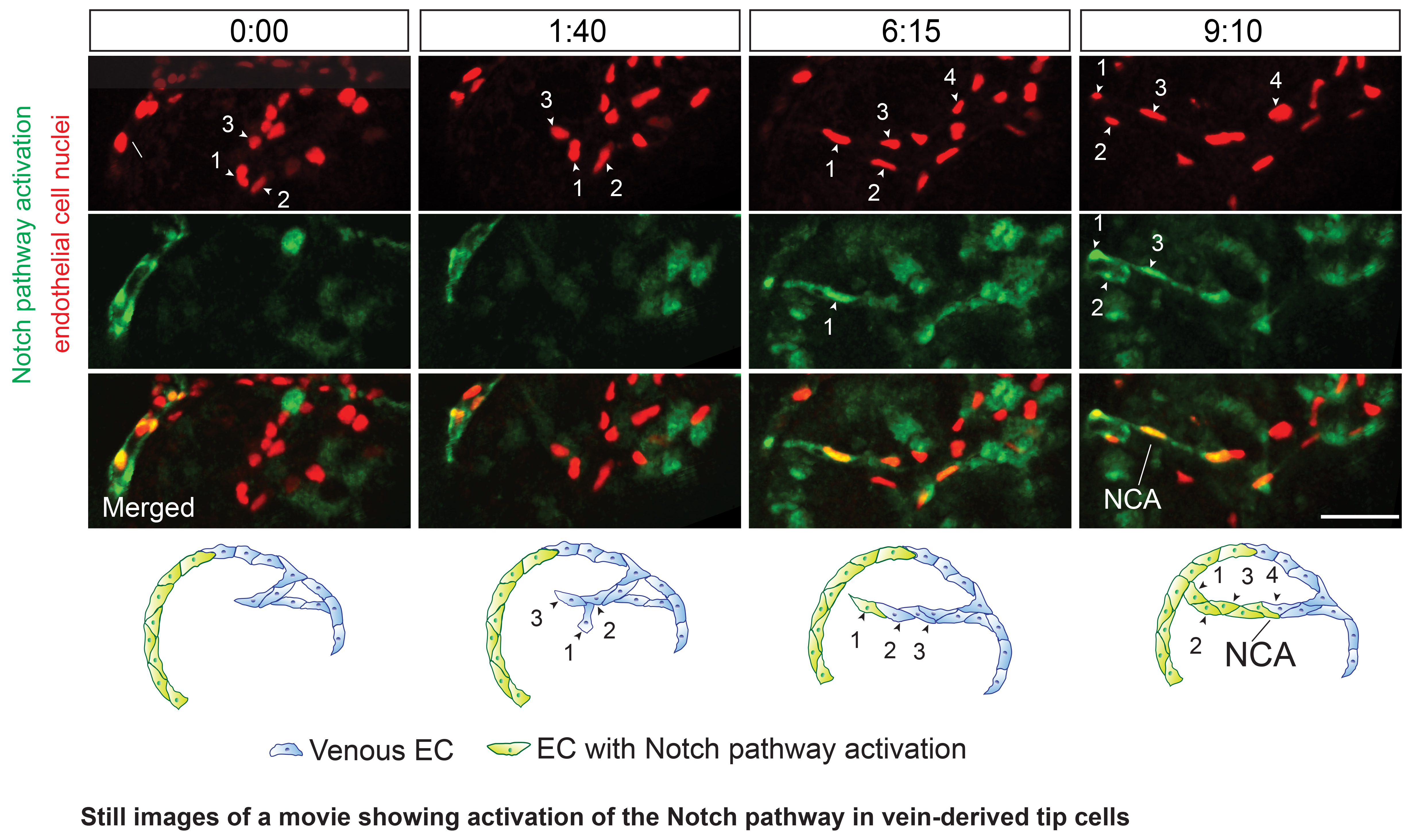
During vascular development, new blood vessels initially sprout out into previously non-vascularized tissues. Subsequently, these blood vessels need to differentiate into different types of blood vessels, such as arteries and veins in order to fulfill different functions. Arteries, for instance, are supported by a layer of smooth muscle cells that help them to sustain the high pressure of arterial blood. Veins, by contrast, contain valves that prevent the backflow of blood. One interest of the laboratory is to understand how arteries are forming. We could previously show, using time-lapse imaging of the regenerating zebrafish fin, that newly forming arteries are generated from pre-existing veins.
We also found that these vein-derived cells show a specific migratory behavior that allows them to connect to arteries that are already present in the tissue. One key factor controlling this migration is the chemokine receptor cxcr4. We went on to show that the Notch signaling pathway, initially implicated in restricting new blood vessel sprouting, is important for regulating cxcr4 expression. Therefore, our results shed light on the intricate interplay of two genetic pathways in coordinating endothelial cell migration during the initial phases of artery formation.
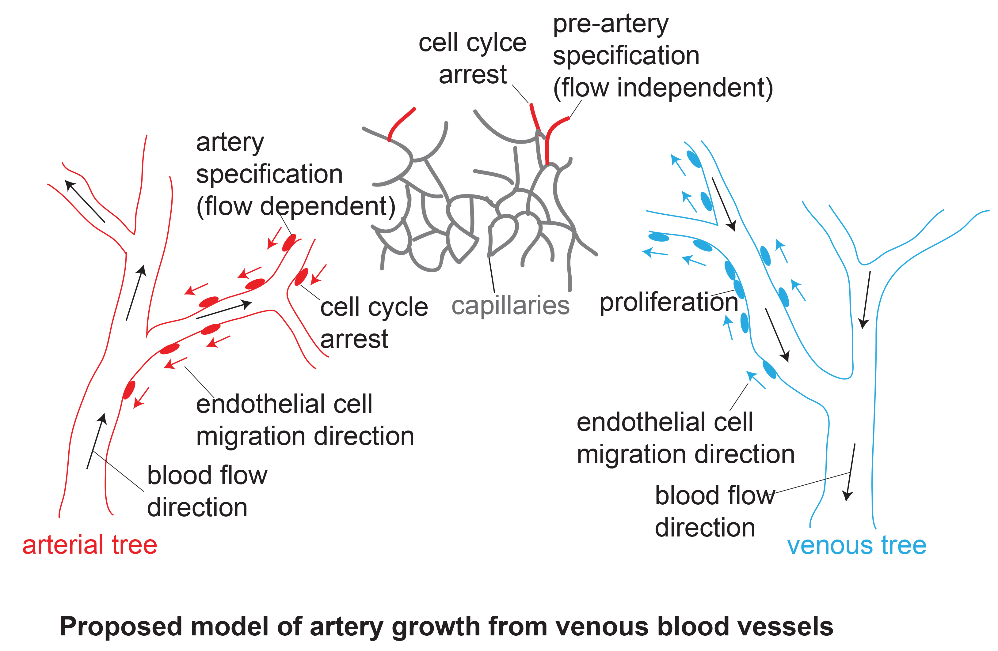
These findings have implications for our understanding concerning the formation of hierarchically patterned blood vessel trees. The vasculature is not a blind ending tree, but rather consists of two trees, an arterial and a venous tree. These are connected at their tips, so that blood can flow between them. The differences in growth direction between veins and arteries that we observed now suggest that veins grow from their bigger stems out towards the branches, just as we expect from a tree. By contrast, arterial trees would grow in the opposite direction, with new cells -coming from veins- being added at the tips of the small branches, from where they would grow towards the larger stem. Of interest, blood flows in the opposite direction through the arterial and venous trees: in the arterial tree, it flows from the bigger stem into the smaller branches, while for the venous side, the small branches collect the blood and transport it to the larger stems. In the future, we want to understand how endothelial cells coordinate these distinct growth directions while at the same time undergoing artery differentiation.
Publications
Siekmann A.F. and Red-Horse K., (2019). Veins and Arteries build hierarchical branching patterns differently: Bottom-up versus Top-down. Bioessays, 41(3):e1800198.
Hasan S.S., Tsaryk R., Lange M., Wisniewski L., Moore J.C., Lawson N.D., Wojciechowska K., Schnittler H., Siekmann A.F. (2017). Endothelial Notch signalling limits angiogenesis via control of artery formation. Nature Cell Biology, 19(8):928-940.
Xu C., Hasan S.S., Schmidt, I., Rocha, S.F., Pitulescu, M.E., Bussmann, J., Meyen, D., Raz, E., Adams, R.H., Siekmann, A.F. (2014). Arteries are formed by vein-derived endothelial tip cells. Nature Communications, 5, 5758.
Siekmann A.F., Affolter M., Belting H.G. (2013). The tip cell concept 10 years after: New players tune in for a common theme. Experimental Cell Research, 319(9):1255-63.
Bussmann J., Wolfe S.A., Siekmann A.F. (2011). Arterial-venous network formation during brain vascularization involves hemodynamic regulation of chemokine signaling. Development, 138 (9):1717-1726.