Welcome to LSPFI
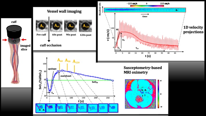
(figure courtesy of Alessandra Caporale).
Research of the Laboratory for Structural, Physiologic and Functional Imaging (LSPFI) is aimed at quantitatively characterizing tissue properties and their relationship to physiology and function by means of spatially resolved magnetic resonance in humans. The laboratory is almost entirely funded by grants from the National Institutes of Health and involves intra- and extramural collaborations with a wide range of specialties, including neurology, psychiatry, cardiology, endocrinology, neonatology, hematology, craniofacial surgery, and orthopaedics.
LSFPI is currently focused on development of novel image-based quantitative methods for the study of tissue oxygen metabolism at enhanced temporal and spatial resolution, and their application to the study of degenerative and acquired vascular disease of the brain and other organ systems. A related area of concentration is development of technology for the evaluation of preclinical vascular disease allowing quantification of markers of endothelial dysfunction in the systemic circulation. Applications of these methods currently in progress target the peripheral, central and neurovascular system in subjects exposed to e-cigarette aerosol inhalation, subjects affected by various lifestyle related conditions, and in patients with sleep disorders. Other ongoing projects target development of new techniques for the study of bone matrix and mineral properties applied to the evaluation and treatment of degenerative bone disease, the conception of methods for constructing cranial models as surgical aids, and assessment of metabolic and degenerative skeletal disorders by means of image-based computational biomechanics with the longer-term goal of creating orthopedic applications, including patient-specific hip fracture prediction involving generation of high-resolution 3D anatomical models.