Breast PET
Principal Investigator: Suleman Surti
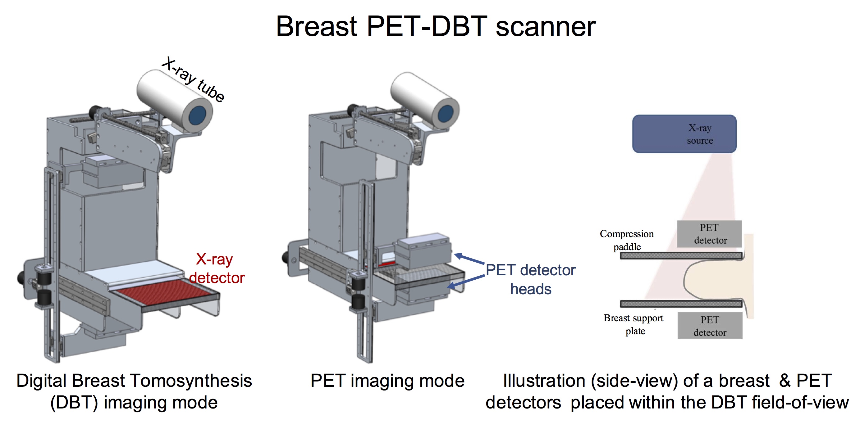
Commercial whole-body PET/CT scanners are limited in their performance when imaging small (< 2 cm in size), early stage tumors or for detecting small changes in tumor uptake. Dedicated breast PET scanners, on the other hand, are geared towards lesion detection (patient screening) and do not emphasize the quantitative imaging capability that is needed for personalized therapy. Our goal is to develop a dedicated, quantitative breast PET scanner that is time-of-flight capable (300 – 400ps) and has higher spatial resolution. The scanner will also be fully integrated with a next-generation digital breast tomosynthesis to generate fully corrected and co-registered PET-DBT images. The multi-modality scanner is expected to benefit early-stage primary tumor characterization, in diagnostic imaging of patients with suspicious, early-stage cancer, as well as in assessing tumor response to therapy.

Selected Presentations and Publications
- Krishnamoorthy S, Vent T, Barufaldi B, Maidment ADA, Karp JS, Surti S. Attenuation correction in a combined, single-gantry breast PET-Tomosynthesis scanner. 2018 IEEE Nuclear Science Symposium and Medical Imaging Conference, Sydney, Australia. M-03-346.
-
Krishnamoorthy S, LeGeyt B, Werner ME, Kaul M, Newcomer FM, Karp JS, Surti S. Design and performance of a high spatial resolution time-of-flight PET detector. IEEE Trans Nucl Sci, vol. 61, pp. 1092-1098, 2014. [Design and Performance of a High Spatial Resolution]
-
Lee E, Werner ME, Karp JS, Surti S. Design optimization of a TOF breast PET scanner. IEEE Trans Nucl Sci, vol. 60, pp. 1645-1652, 2013. [Design Optimization of a Time-Of-Flight]
-
Krishnamoorthy S, Morales E, Ashmanskas WJ, Mayer G, Hu R-W, LeGeyt BC, Karp JS, Surti S. A modular waveform-sampling data acquisition system for time-of-flight PET. 2018 IEEE Nuclear Science Symposium and Medical Imaging Conference, Sydney, Australia. M-07-152.


