PennPET Explorer
Principal Investigator: Joel S. Karp
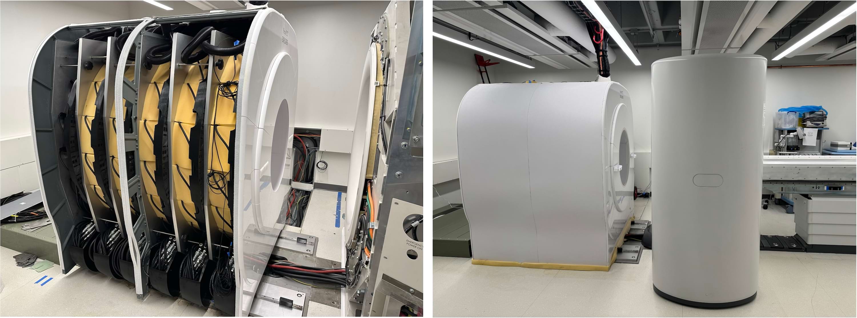
The PennPET Explorer, with an axial field-of-view of 142 cm and coupled to a spectral-CT scanner, is dedicated for research imaging in the Stemmler B22 Imaging facility, and used for total-body human research and large animal protocols. Operation and scheduling is overseen by the PET Center. For further details contact Joel Karp (device capabilities), Austin Pantel (clinical research applications), Stephen McDonald (facility operations), or Erin Schubert (scheduling for scans and radiotracers).
The PennPET Explorer is a whole-body imager that can image the major body organs simultaneously with higher sensitivity than that of commercial devices. The device is scalable in axial length and was initially tested in prototype form with 3 rings and axial field-of-view (AFOV) of 64 cm. The technology of the PennPET Explorer is based on the Philips silicon photomultiplier, and the system achieves a time-of-flight (TOF) resolution of 250 ps and a spatial resolution of 4.0 mm (FWHM) (Karp et al. 2019). We completed a series of pilot human imaging studies with each study designed to test specific capabilities of the device. We demonstrated the ability to scan for shorter duration or, alternatively with less activity, without a compromise in image quality. In clinical patients, the PennPET Explorer better delineates extent of FDG-avid disease. Delayed images, up to 10 half-lives with FDG, reveal biological insight and support the ability to track biologic processes over time. Dynamic imaging studies capture image-derived input functions for kinetic modeling applications. Additional studies with experimental research radiotracers illustrate the benefits from the combination of large axial coverage and high sensitivity.
In 2023, the PennPET Explorer has been expanded from 5 to 6 rings (resulting in a 142 cm AFOV; Dai et al. 2023). The CT was also upgraded to a Phillips IQon Spectral CT.
|
Video of a dynamic FDG study performed on the PennPET Explorer while it was configured with 3 rings (64 cm AFOV). |
This study illustrates the ability to capture fast kinetics at the start of the 1-hour dynamic PET, starting with the bolus injection, and plots the time-activity curves of the major organs over the course of the imaging session. |
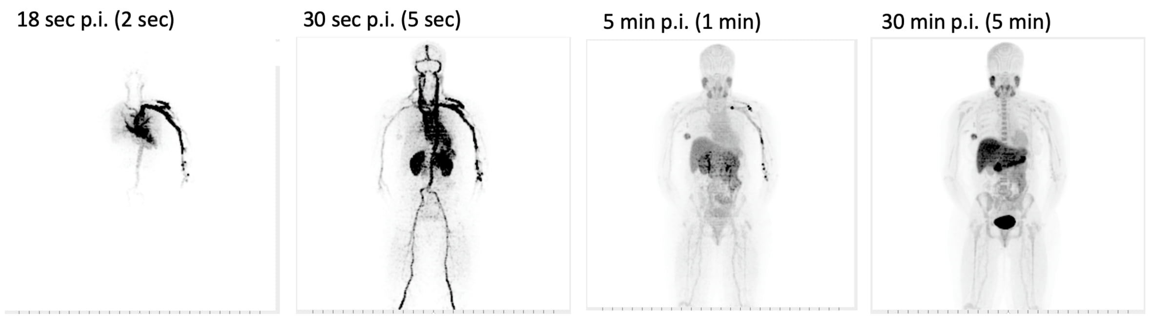
Depending on how the study is designed and the kinetics of the tracer, the dynamic frames can have varying durations, as shown in this 18F-fluoroglutamine study.