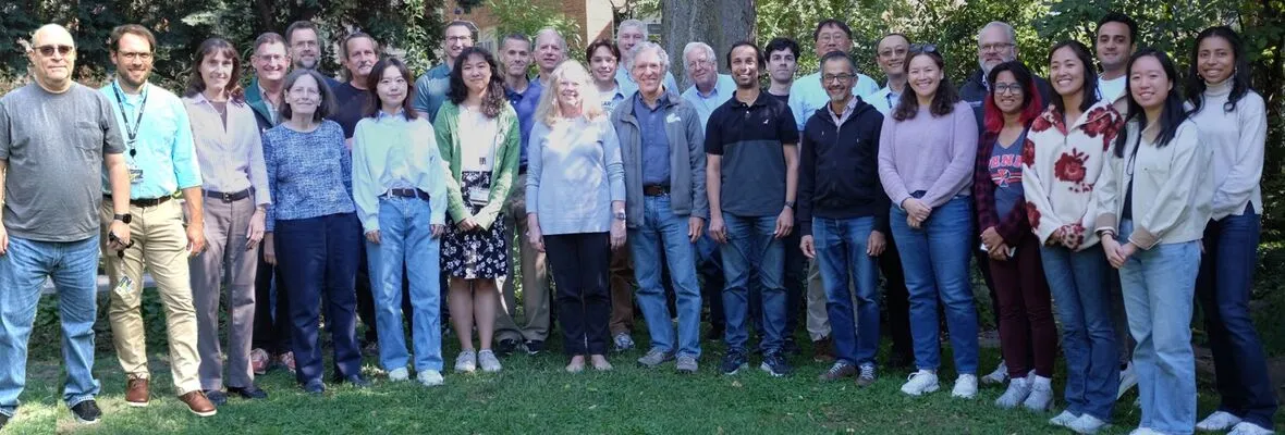
Current personnel
Faculty
-
Read More about Joel Karp, Professor
Joel Karp, Professor
Physics and Instrumentation Group Chief
joelkarp@pennmedicine.upenn.edu
Dr. Karp is Professor of Radiologic Physics in the Department of Radiology, and in the Department of Physics & Astronomy at the University of Pennsylvania. He is Chief of the Physics and Instrumentation Research Group in Radiology and directs Nuclear Medicine/PET Physics and QC in the clinic, as well as the Small Animal Imaging Facility Nuclear Medicine (PET/SPECT/CT) core. He received his PhD in nuclear physics from MIT in 1980 and joined the faculty at Penn in 1983, and since then his research has focused on investigations to improve and characterize the performance of PET technology, including front-end electronics, detector design, data correction techniques, and 3D image reconstruction algorithms. This work has resulted in development of fully 3D PET scanners and innovative imaging systems based on various scintillation detectors, and some of these concepts have been implemented commercially for human and animal imaging. Dr. Karp has developed systems for time-of-flight (TOF) imaging, and his work with industry led to adoption of TOF in modern PET/CT scanners. Dr. Karp is currently leading the team at Penn in the development of the large axial field-of-view PennPET Explorer instrument, which will enable new opportunities for both clinical and research investigations.
-
Read More about Scott Metzler, Research Professor
Scott Metzler, Research Professor
SPECT system design, Small Animal Imaging, Image Reconstruction
Email Scott Metzler, Research Professor
Scott Metzler, Research Professor
SPECT system design, Small Animal Imaging, Image Reconstruction
metzler@upenn.edu
Projects: SPECT system design, Small Animal Imaging, Image Reconstruction
Dr. Metzler is a Research Professor of Radiology. His research focuses on the development and characterization of Single Photon Emission Computed Tomography (SPECT) systems and collimation as well as unique uses of PET scanners, such as the acquisition of supersampled data in PET. He also develops calibration techniques and reconstruction software for these systems.
Dr. Metzler completed his BS in physics at Pennsylvania State University and PhD in physics at University of Pennsylvania. He completed his first post-doctoral training in high-energy physics at California Institute of Technology on the BaBar experiment, which was looking for and found CP violation in B-quark mesons. Dr. Metzler then completed his second post-doctoral training in medical imaging at Duke University and joined the faculty as a research assistant professor. Dr. Metzler joined the Department of Radiology in 2004.
Dr. Metzler's instrumentation efforts are currently geared towards cardiology. He is the principal investigator of the C-SPECT project, which aims to replace traditional myocardial SPECT imaging with a stationary, upright system. An important goal of this project is to measure the flow of blood to different regions of the heart muscle and compare that result with PET, which is currently the gold standard for that measurement, but suffers from substantially higher costs and lower availability. This effort is complemented with parallel developments in small-animal imaging, where it is possible to study the molecular and genetic mechanisms that affect cardiac function. Dr. Metzler is also involved in imaging research for the assessment of Peripheral Arterial Disease with colleagues at University of Illinois and Yale University.
-
Read More about Samuel Matej, Research Professor
Samuel Matej, Research Professor
Image Reconstruction, PET Image Analysis
Email Samuel Matej, Research Professor
Samuel Matej, Research Professor
Image Reconstruction, PET Image Analysis
matej@pennmedicine.upenn.edu
Projects: Image Reconstruction, PET Image Analysis
Dr. Matej is Research Associate Professor of Radiology in the Department of Radiology at the University of Pennsylvania. He received his M.Sc. in computer science from Slovak Technical University in 1983, Ph.D. in measurement science from Slovak Academy of Sciences in 1988, and joined University of Pennsylvania in 1991. He has been active in the research field of tomographic image reconstruction methods for about 35 years, with the main focus directed towards research, development, and investigation of clinically practical fully three-dimensional PET reconstruction methods and tools supporting quantitative PET imaging. These include Fourier-based techniques and novel TOF data partitioning approaches allowing very efficient implementation of analytic and iterative fully 3D reconstruction algorithms. His overall research philosophy has been to develop approaches not only of high scientific quality and novelty, but at the same time approaches that are reliably applied to acquired PET data and that are robust and practical for routine clinical employment. Success of this philosophy has been demonstrated by successful applications of his research results in clinic within our and other institutions, and implementation of the developed reconstruction algorithms and tools on commercial PET systems which are now in routine clinical use. Dr. Matej's current research involvements include TOF reconstruction and data correction approaches for quantitative static and 4D dynamic reconstructions for conventional and long axial FOV (PennPET Explorer) whole-body scanners, as well as for specialized instruments such as dual-panel breast scanner (B-PET), developed within our team at Penn.
-
Read More about Suleman Surti, Research Professor
Suleman Surti, Research Professor
PET System Design, PET Image Analysis, Monte-Carlo modeling
Email Suleman Surti, Research Professor
Suleman Surti, Research Professor
PET System Design, PET Image Analysis, Monte-Carlo modeling
surti@pennmedicine.upenn.edu
Projects: PET System Design, PET Image Analysis, Monte-Carlo modeling
Dr Surti’s general research interest is in PET imaging, ranging from detector and system development through data corrections to image analysis and imaging protocol optimization. His current research projects involve the development of a dedicated breast PET/DBT (digital breast tomosynthesis) system, new scatter correction methods, task based image optimization for modern PET/CT systems, and new PET system design and development.
Dr Surti has been involved in work related to new detector designs with improved spatial and timing performance, design of new PET systems with long axial FOV, as well as improved spatial and timing resolution, and significant experience in analyzing improved imaging performance with TOF PET.
Dr Surti obtained his PhD in Physics at the University of Pennsylvania in 2000 followed by postdoctoral research work also at Penn through 2003.
-
Read More about Min Gao, Research Associate
Min Gao, Research Associate
System Design, PennPET, Brain PET
Min.Gao@Pennmedicine.upenn.edu
Dr. Gao works on novel system design and imaging including a cost-effective total-body PET scanner based on the PennPET Explorer and a dedicated high-performance Brain PET scanner. Her research includes experiments with the PennPET Explorer system, designs and evaluations of advanced detectors and scanners, and lesion detectability studies to understand how instrumental performance influences clinical applications.
Previously during her Ph.D., she developed an all-digital PET dual-head system for in-beam, beam-on range monitoring in proton therapy. She earned her Ph.D. and B.S. in Biomedical Engineering at the Huazhong University of Science and Technology in 2022 and 2017, respectively, and joined the University of Pennsylvania in 2022.
-
Read More about Liz Li, Instructor
Liz Li, Instructor
PET kinetic modeling, image quantification and analysis
Liz.Li@Pennmedicine.upenn.edu
Liz joined the Physics and Instrumentation Group after working as a postdoctoral fellow at UC Davis, where she also earned her Ph.D. in Biomedical Engineering. Liz has a B.S. in Mathematics from UCLA and an M.S. in Biomedical Imaging from UCSF. Prior to joining the EXPLORER Consortium at UC Davis as a Ph.D. student, she worked in industry where she was involved in various clinical imaging projects.
Liz is interested in quantitative PET imaging and analysis, with a focus on PET kinetic modeling and the use of an image-derived input function. Additionally, she is interested in how the increased sensitivity of long axial field of view PET scanners like the PennPET Explorer can be leveraged to improve upon clinical assessments and quantification. She also provides user support in the Nuclear Medicine Advanced Analysis Lab.
In her free time, she enjoys crafting, spending time outdoors with friends and her cat Willie Jack, as well as reading sci-fi novels.
Adjunct Faculty
-
Read More about Steve Moore, Adjunct Associate Professor
Steve Moore, Adjunct Associate Professor
PET and SPECT, Small Animal Imaging
Email Steve Moore, Adjunct Associate Professor
Steve Moore, Adjunct Associate Professor
PET and SPECT, Small Animal Imaging
scmoore1@pennmedicine.upenn.edu
Projects: PET and SPECT, Small Animal Imaging
Dr. Moore is currently working with Dr. Metzler and colleagues on developing and optimizing new systems and kinetic-modeling techniques for quantitative imaging of myocardial blood flow and flow reserve in various research applications. He is also working with Drs. Karp, Metzler, and other colleagues on new approaches for simultaneous dual-radionuclide PET imaging, both for preclinical PET systems and for the PennPET Explorer total-body PET scanner.
During a post-doctoral position in Radiology at Brigham and Women's Hospital in Boston, and through successive faculty positions at Harvard Medical School, Dr. Moore worked on research projects related to prospectively gated cardiac CT, as well as on task-based optimization of systems for improved nuclear medicine imaging and image-based quantitation. From 1986 to 1992, while on the faculty in Biomedical Engineering at the Worcester Polytechnic Institute, he advised numerous graduate and undergraduate students on their research projects, continued his research in nuclear medicine, and gained experience in magnetic resonance imaging research. Dr. Moore then returned to the Joint Program in Nuclear Medicine at Harvard Medical School, eventually to become the Director of Nuclear Medicine Physics at Brigham and Women's Hospital, where his research was based on improving the acquisition, reconstruction, and extraction of quantitative information from emission tomographic images; in that capacity, he was principal investigator on a long-term NIH R01 grant, and he also developed a multiple-pinhole research micro-SPECT imaging system with additional support from NIH and industry.
Dr. Moore earned a B.S. in physics from Bucknell University in 1972, and a Ph.D. in experimental high-energy physics from Brandeis University in 1978. After a short period working in industry, he received additional training as a post-doctoral research associate in Radiologic Physics and Nuclear Medicine at Brigham and Women's Hospital and Harvard Medical School.
Dr. Moore is now semi-retired; he and his wife enjoy traveling, babysitting for their grandchildren in Philadelphia, and spending much of the summer and many weekends at their camp on a lake in the Adirondacks. During the school year, Dr. Moore has been volunteering as a tutor for underserved middle-school students in math and science in the after-school program at the North Light Community Center. He continues to pursue a long-time interest in music by attending many concerts, and by practicing on his 110-year-old Steinway piano.
Research Staff
-
Read More about Bill Ashmanskas
Bill Ashmanskas
Senior Research Investigator
ashmansk@hep.upenn.edu
Bill Ashmanskas splits his time as a Sr. Research Investigator in the Department of Radiology and a Senior Lecturer in the Department of Physics. His background as a high-energy physicist and experience with electronics gained at Fermi National Accelerator Lab translated to the development of PET electronics for time-of-flight PET imaging. He is currently responsible for the development of digital electronics for the B-PET project.
-
Read More about Bing Dai
Bing Dai
Senior Research Investigator
bing.dai@pennmedicine.upenn.edu
Projects: PennPET Explorer
Dr. Dai's broad research interests include programming, electronics, optics, computer vision, image processing, nanofabrication, etc.., with a special interest in tackling interdisciplinary challenges with holistic solutions emphasizing the close interaction between hardware innovation and software advancement. He is currently responsible for the firmware development for the PennPET Explorer total body imager.
He was the lead scientist for developing hardware control solutions for open source laser scanning systems at the Laboratory for Optical and Computational Instrumentation (LOCI) at the University of Wisconsin–Madison from 2015 to 2019, and an advisory engineer providing computational patterning solutions for five generations of IBM semiconductor technologies, at IBM, New York, from 2011 to 2014. Before that, he developed a super high resolution (sub 10 nm) x-ray microscopy for non-destructively imaging of nanoscale structures at Stanford / SLAC.
He received his PhD degree in applied physics and his MS degree in electrical engineering from Stanford University in 2010 and 2009, respectively, and his BS degree in physics from Peking University, Beijing, China, in 2005.
He is an avid reader, hiker, and traveler, and likes exploring history, culture, and architecture in his spare time.
-
Read More about Margaret Daube-Witherspoon
Margaret Daube-Witherspoon
Senior Research Investigator
daubewit@pennmedicine.upenn.edu
Projects: Image Reconstruction, PET Image Analysis and data quantification
Dr. Daube-Witherspoon's research encompasses all aspects of quantitative PET imaging, from testing/optimizing reconstruction algorithms to corrections methodology to image analysis. She is currently focused on long axial FOV PET scanner characterization of both intrinsic performance and applications unique to these systems.
From 1983-1986, Dr. Daube-Witherspoon worked in the Department of Radiology at the University of Pennsylvania with Gerd Muehllehner. From 1986-1998, she was at the National Institutes of Health, first in the Nuclear Medicine Department and then in the Positron Emission Tomography Department.
She received her B.A. in Physics from Swarthmore College in 1978 and her Ph.D. in Physics from the University of Wisconsin - Madison in 1983. Her Ph.D. thesis work involved developing new PET radiotracers for myocardial perfusion imaging, from accelerator target development to human biodistribution studies.
-
Read More about Srilalan Krishnamoorthy
Srilalan Krishnamoorthy
Senior Research Investigator
srilalan@pennmedicine.upenn.edu
Projects: PET System Design
Dr. Krishnamoorthy has experience on several aspects related to the development of instrumentation for PET scanners and is working on the development of a high spatial resolution, time-of-flight capable PET scanner for dedicated breast imaging.
Dr. Krishnamoorthy's research before joining Penn focused on the design, development and optimization of several scintillation and semiconductor detectors for gamma-ray imaging.
He received both his Master's and Doctoral degrees (Stony Brook University) in Biomedical Engineering with a focus on Medical Imaging Physics.
-
Read More about Matt Werner
Matt Werner
Senior Software Architect
matt.werner@uphs.upenn.edu
Projects: PennPET Explorer, Image Reconstruction, Scanner Calibration
Matt is interested in hardware calibrations, data corrections, and image reconstruction architectures related to multi-ring long axial field-of-view PET scanners.
Matt has nearly 20 years of experience in developing algorithms and software related to scanner efficiency and timing calibration, scatter correction, and 3D image reconstruction. Before that, Matt worked in the aerospace industry developing simulations, 3D visualizations, and performance analyses of missile defense and satellite surveillance systems.
Matt has a BS in Mechanical Engineering from RPI, a MS in Mechanical Engineering from Caltech and a MS in Computer Science from Penn.
-
Read More about Michael Geagan
Michael Geagan
Senior System Design Engineer
mgeagan@pennmedicine.upenn.edu
Projects: PennPET Explorer
Michael is responsible for the mechanical and system design of the PennPET Explorer total body imager. He has worked as the Director of Engineering and Advanced Development at Philips Medical Systems PET Imaging Group. Michael has a BA from the University of Pennsylvania.
-
Read More about Mike Parma
Mike Parma
Senior Software Architect
parma@pennmedicine.upenn.edu
Projects: PennPET Explorer
Mike Parma works on software architecture, software development, hardware support, and 510k support for the PennPET Explorer project. He has over 30 years of software development experience in the medical device industry, including both PET and CT. Mike Parma has a BS in Computer Engineering, 1985 from Case Western Reserve University, Cleveland OH.
-
Read More about Dale Stentz
Dale Stentz
Research Staff
dstentz@pennmedicine.upenn.edu
Projects: C-SPECT
Dr. Stentz works on designing and building the Cardiac SPECT Imaging. He previously worked on the CDF experiment, a high-energy physics experiment at Fermilab. He completed hisPhD at Northwestern in physics.
-
Read More about Emmanuel Morales-Negron
Emmanuel Morales-Negron
Tech Instumentation
Emmanuel Morales-Negron
Tech Instumentation
Projects: B-PET
Emmanuel has been working with the High Energy Physics Instrumentation group at the University of Pennsylvania developing a quad-channel amplifier for fast timing application. He is also working on the development of custom electronics being developed for a dedicated breast PET scanner.
-
Read More about Amy Perkins
Amy Perkins
Technical Project Manager, Philips Healthcare
Amy Perkins
Technical Project Manager, Philips Healthcare
Projects: PennPET Explorer, Spectral CT
As a Philips research physicist, Dr. Perkins works on-site at the University of Pennsylvania collaborating with the UPenn PET group to build the PennPET Explorer and investigate the benefits of long axial-field-of-view scanners. Amy also collaborates with Dr. Peter Noel at UPenn on spectral CT imaging research.
Amy has participated in feasibility testing and the building of PET prototypes, including a LaBr3-based scanner, the A-PET animal system, and a pixelated NaI(Tl) prototype. She has performed studies with clinical data to evaluate the clinical impact of time-of-flight imaging, as well as investigated the effect of different imaging corrections on quantitative accuracy.
Dr. Perkins received her PhD in physics from Brown University
-
Read More about Anthony Young
Anthony Young
Research Specialist
Anthony Young
Research Specialist
Projects: PET Image Analysis
Anthony performs PET/CT and MRI image analysis and kinetic modeling on new and existing PET radiotracers, studying cancer, infection, addiction, and neurodegeneration. He also designed and implemented an application for measurement of 3D spinal curvature, coauthoring the patent "Systems and Methods for Modeling Spines and Treating Spines based on Spine Models" (US Patent Appl. 20190099221). He graduated from Drexel University with a B.S. in Biomedical Engineering.
Clinical Physics
-
Read More about Janet Reddin
Janet Reddin
Senior Research Investigator
Janet Reddin
Senior Research Investigator
Dr. Reddin is a Sr. Research Investigator and contributes to the support of the clinical instruments in Nuclear Medicine, including monthly, quarterly, and annual testing of existing equipment and acceptance testing of new PET/CT and SPECT/CT scanners. She also is a course director of the Nuclear Medicine physics curriculum for Radiology residents and Nuclear Radiology fellows. Since 2005 she has served as a PET Core Lab Physicist at ECOG/ACRIN, qualifying hundreds of PET/CT scanners for participation in quantitative PET research trials. She is also Co-Director of the University’s Nuclear Medicine Advanced Image Analysis Lab (NM AIA).
-
Read More about Josh Scheuermann
Josh Scheuermann
Medical Physicist
Josh Scheuermann
Medical Physicist
Projects: PennPET Explorer, PET Image Analysis, Clinical Support for NucMed and PET Clinics
Josh is mostly involved in supporting the physics needs of the Nuclear Medicine and PET clinics, including internal dosimetry for radionuclide therapies and quality assurance for the Nuclear Medicine and PET cameras. He has gotten involved in the PennPET Explorer project to assist in moving the system into a more clinical operating environment.
Josh started working for the Physics and Instrumentation Group as a junior level physicist in 2006 right out of graduate school. For his first few years, his focus was on image analysis for the ACRIN PET Core Lab and updating and standardizing the quality control programs in the Nuclear Medicine and PET clinics at HUP, PCAM and several satellites. In 2012, Josh began cross-training in Radiation Oncology and has been working as a clinical medical physicist in both Nuclear Medicine and Radiation Oncology since.
Josh graduated from St. Joseph's University in 2004 with a BS in Physics. He then enrolled in the Master of Medical Physics program at University of Pennsylvania and graduate with an MMP degree in 2006.
While not at work, Josh enjoys carpentry, vegetable gardening, traveling to new places and getting outdoors to go backpacking, hiking and camping. He and his wife recently had a son and they are looking forward to continuing their traveling with Vance and imparting their love of the outdoors to him.
-
Read More about Randy Kulp
Randy Kulp
Electrical Engineer
Randy Kulp
Electrical Engineer
Projects: PennPET Explorer, Small Animal Imaging, Clinical Quality Control
Randy works on retrofitting and updating legacy scanners and developing new systems. He has 30 years electrical engineering and prototyping experience. He has also worked as a Philips Medical Systems lab technician troubleshooting and calibrating new PET systems in the lab. Additionally, Randy has field experience in trouble shooting and corrective maintenance of systems, along with setting up new scanners and systems in research labs. Randy has an Electrical Engineering Associates Degree.
-
Read More about Stephen McDonald
Stephen McDonald
Research Specialist
stephen.mcdonald@pennmedicine.upenn.edu
Projects: PennPET Explorer
Stephen is currently performing research on the PennPET Explorer. His main responsibilities are to service and maintain the scanner while running performance and quality tests. He is also working on constructing the rings of the PennPET Explorer to an eventual 6 ring scanner.
He worked as an HVAC apprentice while in college. He has also worked in a fuels lab where he tested the chemical emissions of coal and petroleum coke in Jacksonville.
Stephen studied engineering/physics at Jacksonville University and the University of North Florida.
SAIF
-
Read More about Eric Blankemeyer
Eric Blankemeyer
Technical Director, SAIF Nuclear Medicine Core
eriblank@pennmedicine.upenn.edu
Projects: Small Animal Imaging
Eric is currently, the Technical Director of the Small Animal Imaging Facility. His responsibilities include animal imaging, development of imaging protocols, and maintenance of the PET, SPECT, and CT scanners in the lab. Eric has a BS in Biological Sciences from Rutgers University
Administration
Postdoctoral Fellows
-
Read More about Majid Kazemi Kozani
Majid Kazemi Kozani
Postdoctoral Researcher
Majid.Kazemi@Pennmedicine.upenn.edu
Project: Investigating the correlation between numerical observer and human observer in lesion detection tasks.
Dr. Kazemi Kozani is presently engaged in assessing the task-specific image quality of PennPET Explorer. His research encompasses studies using human and numerical observers to estimate lesion detectability and tumor response to therapy, offering correlational data for numerical observers, including metrics like NEC.
Having earned a B.Sc. in Physics from Shahid Beheshti University, Dr. Kazemi pursued his M.Sc. in Radiation Application Engineering at Amirkabir University of Technology, Tehran. He then received his Ph.D. in Physics (2022) with a focus on "Event Pattern Recognition and Image Reconstruction in Compton Camera for Proton Therapy Monitoring" from Jagiellonian University. Before joining the Physics and Instrumentation Group in 2023, Dr. Kazemi worked as a researcher at the J-PET collaboration at Jagiellonian University, where he contributed to the development of J-PET as a Compton camera for improved patient-specific diagnosis and treatment planning.
Graduate Student Researchers, Scholars
-
Read More about Florence Muller
Florence Muller
Visiting Research Scholar
Florence.Muller@Pennmedicine.upenn.edu
Projects: PennPET Explorer, image-based deep learning applications in PET
Florence obtained a M.Eng. in Mechanical and Materials Engineering from the University of Birmingham (UK) in July 2020 and a M.S. in Biomedical Engineering from Ghent University (Belgium) in July 2022. Since August 2022, she is a Ph.D. student in Biomedical Engineering with the Medical Image and Signal Processing group at Ghent University; she is spending her first year abroad at the University of Pennsylvania as part of the Physics and Instrumentation Group. As a visiting research scholar, she is jointly funded by Fulbright and the Belgian American Educational Foundation.
At UPenn, Florence is currently working on developing deep learning strategies for image denoising (3D and 4D) and PET-CT dose reduction (e.g., CT-less attenuation correction in PET). Her scientific interests include the integration of artificial intelligence methods into image generation and post-processing to obtain more quantitatively accurate PET and CT image for clinical and preclinical applications.
-
Read More about Raheema Demani
Raheema Demani
Graduate Student Researcher
Raheema Demani
Graduate Student Researcher
Project: Evaluating novel imaging markers from dual tracer PET studies
Raheema Damani is a Bioengineering Ph.D. student and a Howard Hughes Medical Institute (HHMI) - Interfaces Fellow at the University of Pennsylvania. She graduated from University of Alabama at Birmingham with a Bachelors in biomedical engineering. Her research interests are in leveraging machine learning algorithms to extract novel imaging markers from dual tracer PET studies.
-
Read More about Shweta Majumder
Shweta Majumder
Graduate Student Researcher
Shweta Majumder
Graduate Student Researcher
Project: Energy-based scatter PET, PET kinetic modeling
Shweta joined the Physics and Instrumentation Group as a second-year PhD student in Bioengineering at the University of Pennsylvania under the supervision of Dr. Suleman Surti. Her research focuses on energy-based scatter correction in PET imaging, particularly within the context of the Penn-PET Explorer. Additionally, she collaborates with Dr. David Mankoff and others on PET kinetic modeling.
Before starting her doctoral studies, Shweta earned a Sc.B. in Engineering Physics from Brown University. She then worked as a research associate at the Yale School of Medicine's Department of Neurology, where her work centered on EEG analysis for tactile perception.
Undergraduate Student Researchers
-
Read More about Jelena Vázquez Delgado
Jelena Vázquez Delgado
PURM Undergraduate Summer Researcher
Jelena Vázquez Delgado
PURM Undergraduate Summer Researcher
Project: FNOS vascular inflammation
Jelena is a Penn undergraduate from Bogotá, Colombia majoring in Bioengineering. She is particularly interested in the intersection between mathematics and biomedical imaging and hopes to pursue a PhD in a related field in the future. In her free time, Jelena enjoys reading, trying new foods, dancing and spending time with family and friends.
-
Read More about Joshua Pascale
Joshua Pascale
Undergraduate Summer Researcher
Joshua Pascale
Undergraduate Summer Researcher
Project: Investigation of thalamic nuclei with 11C-Cfn and partial volume corrections
Joshua is a Penn undergraduate originally from Secaucus, NJ. He is majoring in neuroscience on a pre-med track minoring in chemistry, health care management, and medical sociology. Joshua hopes to attend medical school after graduating from Penn and potentially pursue a career in neurology.
In his free time, Joshua enjoys working out, running, going out with friends, and binging TV shows.
-
Read More about Sophie Lammers
Sophie Lammers
Undergraduate Researcher, SUPERS student
Sophie Lammers
Undergraduate Researcher, SUPERS student
Sophie is a Penn undergraduate originally from Evanston, IL. She is majoring in Physics with a concentration in Biological Science and minoring in Mathematics and Hispanic Studies. Sophie is primarily interested in medical physics and hopes to pursue a PhD in the field after graduating from Penn.
In her free time she enjoys reading fiction novels, knitting, swimming, spending time with friends, and going to Penn sports games as a member of the PennBand.
Group Founder
-
Read More about Gerd Muehllehner
Gerd Muehllehner
Gerd Muehllehner
Projects: PennPET Explorer
After receiving his Ph.D. in Nuclear Physics, Dr. Muehllehner worked for 12 years for Searle Radiographics (now Siemens) developing Nuclear Medicine instrumentation, specifically gamma cameras. His interest in PET was one of the reasons he decided to join a team at the University of Pennsylvania, where he developed PET scanners for more than 10 years. In 1990 his wife, whose background is in finance, and he decided to make the PET scanner, which was developed at the U of Penn, commercially available at their company, UGM Medical Systems. This scanner was marketed for 5 years through General Electric and later through ADAC. During the years at UGM Medical Systems Dr. Muehllehner was involved in a number of important developments. Starting with a NaI(Tl) based scanner with flat crystals, they pioneered the use of curved NaI(Tl) crystals in a commercial system (the C-PET scanner). Together with the U of Penn they built an animal scanner (marketed through Philips as the Mosaic), a dedicated brain scanner (now used at Penn for primate imaging) and finally a whole body PET scanner using GSO as the scintillator (the Allegro scanner). In 1999 UGM Medical Systems merged with ADAC, which itself merged with Philips Medical Systems a year later. After serving as president at UGM Medical Systems and directing the growth of the company from 3 scanners per year to 60 scanners per year, Dr. Muehllehner went back to Advanced Development during his years at ADAC and Philips Medical Systems. Since April 2005 he has been retired.
Dr. Muehllehner was most recently involved in the development of the PennPET Explorer and formed a small company, KAGE Medical, to leverage his previous experience of taking a University-based instrument to the product stage as part of the work in an NIH Academic-Industry Partnership grant along with Dr. Karp. The goal is to enhance both the performance and operational procedures of the PennPET Explorer scanner, along with high reliability and usability that will enable Penn to perform clinical as well as research PET studies. Sadly, Gerd passed away on June 21, 2020. For a brief summary of his accomplishments in Nuclear Medicine, see In Memoriam published in the Journal of Nuclear Medicine (https://jnm.snmjournals.org/content/61/8/19N)


