
Positron Emission Tomography Center (PET Center)
The Department of Radiology PET Center operates four whole-body PET/CT scanners; Philips Gemini TF Ingenuity, Philips Gemini TF BB, Siemens Biograph mCT, and Siemens Biograph Vision. All scanners are located in the Perelman Center for Advanced Medicine. Separate rooms are available for patient preparation and injection of radiopharmaceuticals. In addition, there is a nearby hot lab for preparing and extracting doses and a blood lab for sampling and counting. The Philips Gemini TF BB is located in Radiation Oncology and used primarily for treatment planning, whereas the other scanners are located in Radiology and used for diagnostic clinical imaging. The Philips Gemini TF Ingenuity is shared 50/50 between research and clinical examinations. The radiotracers are provided by the Cyclotron Facility. (PET Center Website).
The PET Center has a number of functions including: system quality control (QC), protocol optimization and development, and the development of and testing of new PET/CT systems. While daily QC is performed by the Nuclear Medicine technologists, system calibration and maintenance is provided by the Physics and Instrumentation Group in the Nuclear Medicine section of Radiology. In addition to providing imaging protocol development and algorithm optimization to improve imaging performance and ensure accurate image quantitation, the clinical physicists in our group also test the implementation of new instruments. In 2005, our PET Center received the first time-of-flight (TOF) PET/CT scanner, the Philips Gemini TF and performed the initial characterization of this system [Surti et al JNM 2007]. The underlying technology of this new TOF scanner was developed collaboratively with our group, and the resulting characterization of the clinical benefits of TOF [Karp et al JNM 2008] helped to establish TOF PET/CT as the standard diagnostic tool. The most recent PET/CT system in the PET Center is the Siemens Biograph Vision, which was installed in October 2018. This system is based on the newest solid-state photosensor technology and offers improved time-of-flight (TOF) resolution of 210 ps. It also has the largest axial field-of-view of 26 cm and the highest sensitivity of 15.6 kcps/MBq. Having the first installation of this new PET/CT system in the USA, our group reported on the performance of this system in [Reddin et al, MIC 2018].
An example of a clinical FDG study with the Siemens Biograph Vision is shown below. The excellent image quality of this large patient with high BMI is due to a combination of the high sensitivity and excellent TOF resolution.
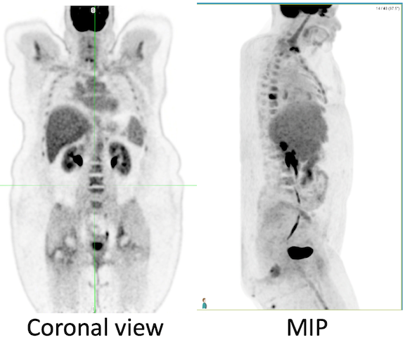
Injection: 15.0 mCi.; Uptake: 54 min
Table speed: 1.4 mm/s, Scan duration: 11 min
Recon: PSF+TOF, 4 iter, 5 subsets, 5 mm Gaus. Voxels: 1.65 x 1.65 x 4mm3