Small Animal Imaging Facility (SAIF)
The Small Animal Imaging Facility (SAIF) at the University of Pennsylvania combines state-of-the-art instrumentation and a nationally recognized staff to assist investigators with a wide range of imaging based experimental approaches. The SAIF is organized into four Imaging Sub-Cores and an animal holding unit. These include MRI/MRS, Nuclear Medicine (PET/SPECT/CT), Optical/Bioluminescence, and Ultrasound. (SAIF Website).
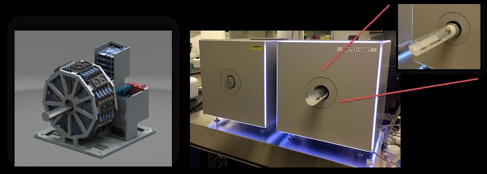
The Nuclear Medicine core, directed by Professor Joel Karp in the Department of Radiology, has recently added the Beta-Cube and X-Cube scanners from Molecubes to their other PET, SPECT and CT equipment. (see Figure below). The Beta-Cube PET scanner is based on a monolithic PET detector and achieves 1-mm3 spatial resolution with high sensitivity. The CT allows for fast, low dose imaging with 85 mm spatial resolution.
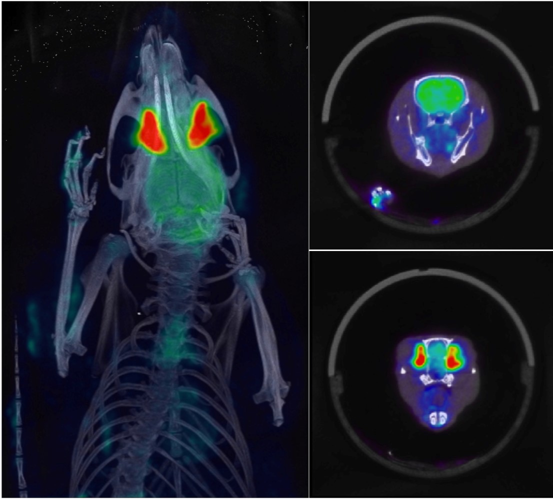
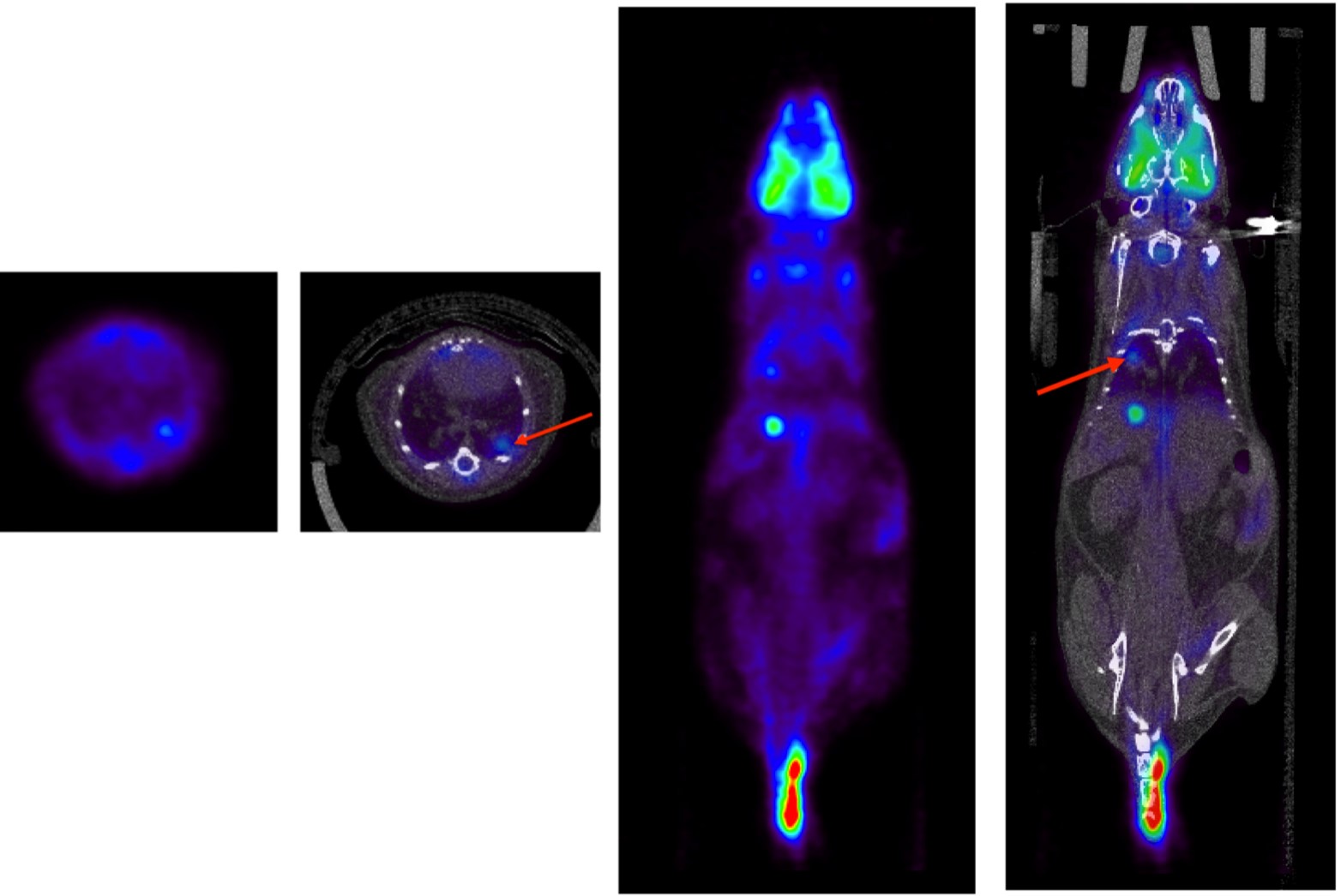
Following the installation at Penn the performance of these instruments was characterized and reported in [Krisnamoorthy PMB 2018 paper]. They are now routinely being used for a wide variety of research studies in both the body and brain of rats and mice. These include studies of cancer metabolism and glutamine uptake, inflammation, and studies of Parkinson’s, Alzheimer’s, hypoxia and ROS biology.
Two examples are shown below.