Laser-scanning Confocal Microscopes
Room B30C Anatomy-Chemistry
Features:
- Zeiss Axio Observer 7 inverted microscope with Definite Focus 3
- ZEN Blue v. 3.5
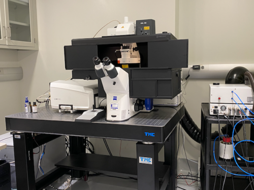
- 6 solid-state visible light lasers: 405 nm, 445 nm, 488 nm, 514 nm, 561 nm, 639 nm
- 2 metal alkali PMTs + 32-channel GaAsP detector for fast spectral characterization
- 730 nm laser + two dedicated NIR detectors for imaging dyes such as Cy7, Alexa Fluor 750, etc.
- motorized XY stage + AI sample finder for automated sample location and scanning
- incubation enclosure for live cell imaging
- Objective lenses: 5x/0.16 NA; 10x/0.45 NA; 20x/0.8 NA; 40x/1.1 NA water imm; 63x/1.4 NA oil imm.
Room 1175A BRB
Features: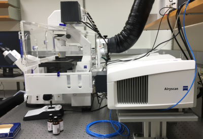
- Zeiss Axio Observer inverted microscope
- ZEN v2.3 software for acquisition
- Excitation laser lines: 405, 458, 488, 514, 561, 633 nm
- Objectives: 10x/0.3 dry, 20x/0.8 dry, 25x/0.8 Imm, 40x/1.2 W, and 63x/1.4 Oil
- Three spectral detection channels: 2 metal alkali PMT + 1 GaAsP detector
- Airyscan module with FAST option for enhanced resolution, speed, and sensitivity
- Environmental chamber and motorized x-y stage for live imaging and tiling applications
Best suited for: confocal imaging of fixed or live samples, including those requiring incubation; FRAP, photoactivation, or photoconversion in live cells or organisms; Airyscan module provides 1.7x increase in resolution; FAST module provides 4x increase in speed.
For new users of this confocal: a handy quick guide to the ZEN software interface.
Room 1-126C Smilow
Features 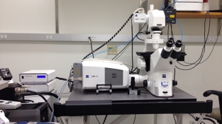
- Zeiss AxioObserver inverted microscope
- ZEN 2012 software for acquisition
- Excitation laser lines: 405, 458, 488, 514, 561, 594, 633 nm
- Objectives: 10x/0.45 dry; 20x/0.8 dry; 25x/0.8 Imm; 40x/1.1 W; 63x/1.4 Oil
- Two metal alkali PMTs and one 32-channel linear array PMT
- Motorized x-y stage for live imaging and tiling applications
Best suited for: confocal imaging of fixed samples or live samples that do not require incubation; FRAP, photoactivation, or photoconversion in live cells or organisms; FRET; spectral characterization and linear unmixing of unorthodox dye combinations
For new users of this confocal: a handy guide to the ZEN software interface
Room 1-126D Smilow
Features:
- DMi8 inverted microscope
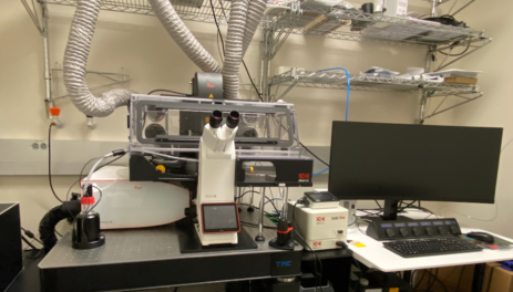
- LAS X software for acquisition
- Extended tunable WLL excitation from 485-790 nm plus fixed 405 nm laser
- Objectives: 10x/0.4 dry; 20x/0.75 dry; 25x/0.95 W; 40x/1.10 W; 40x/1.3 Oil; 63x/1.4 Oil
- Four Power HyD S detectors
- Okolab incubation enclosure
- Motorized x-y stage + Leica's Navigator for tile scans
- FRAP and FRET wizards
- Lightning for deconvolution and TauSense for lifetime-based spectral separation
Best suited for: confocal imaging of fixed or live samples, including those requiring incubation; FRAP, photoactivation, or photoconversion in live cells or organisms; FRET
Room B30A Anatomy-Chemistry
Features: 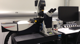
- Leica DMI 6000 inverted microscope
- Pulsed white-light laser for tunable excitation from 470 to 670 nm
- CW laser lines for conventional confocal imaging: 405, 458, 488, 514 nm
- Objectives: 10x/0.4 dry; 20x/0.75 Imm; 40x/1.3 Oil; 63x/1.4 Oil; 100x/1.4 Oil
- High-speed resonant scanner for live-cell imaging
- 3 HyD and 2 PMT detectors
- Two depletion lasers (592 and 660 nm) for super-resolution imaging of green and red dyes
- Tokai Hit stagetop incubator
- Motorized xy stage
- LAS X software for acquisition; Huygens Professional software for deconvolution
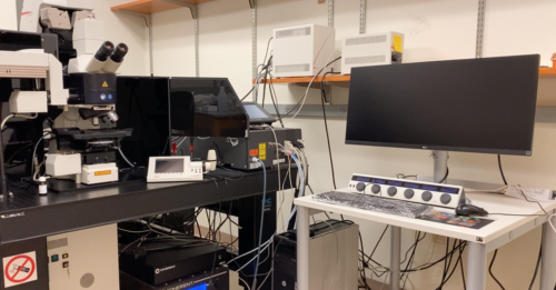
Room 514A CRB
Features:
- Visible laser lines (all diode or DPSS lasers): 405, 488, 514, 552, 638
- Coherent Chameleon Vision II + MPX wavelength extender for tunable excitation from 740 — 1300 nm
- Leica DM 6000 upright microscope
- Objectives: 10x/0.3 dry; 20x/0.75 Imm; 40x/1.3Oil; 63x/1.4 Oil; 25x/1.0 W motCORR available upon request
- High-speed resonant scanner for live-cell imaging
- 2 HyD and 3 PMT confocal detectors
- HyD and PMT dual NDDs for reflected light (fluorescence or SHG)
- PMT NDD for transmitted light (SHG)
- Motorized xy stage
- LAS X software for acquisition