New User
Consultation
All New Users are required to email for a consultation (ULTRA_SERV@lists.upenn.edu). Once you send the email with your request, the EMRL staff will set up a time for a detailed discussion about the project's feasibility, pricing, and goals.
Returning User
If you have already finished consulting with the EMRL staff and would like to drop off samples or sign up for the equipment, proceed to the iLab website.
TEM In Brief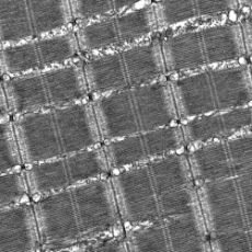
The Transmission Electron Microscope is ideal for imaging various samples from life sciences, medical, and biological to Industrial applications. TEMs provide topographical, morphological, and compositional information.
The images allow researchers to view samples on a molecular level, allowing them to analyze structure and texture.
Negative staining for TEM is widely used to visualize at high resolution several biological samples (e.g., viruses, protein molecules, macromolecular assemblies and fibrils, liposomes, exosomes, synthetic DNA arrays, and polymer solutions)
| Internal | Academic (External) | Corporate (External) | |
|---|---|---|---|
| Critical Point Dryer | $20.00 | $32.60 | $40.00 |
| Duplicate block for post-embedding IEM | $225.00 | $366.75 | $450.00 |
| Duplicate block for TEM analysis | $175.00 | $285.25 | $350.00 |
| Each additional antibody | $200.00 | $326.00 | $400.00 |
| Talos L120C images performed by staff | $150.00 | $244.50 | $300.00 |
| Talos L120C training | $200.00 | $203.75 | $250.00 |
| Talos L120C per hour | $50.00 | $81.50 | $100.00 |
| Post-Embedded IEM | $475.00 | $774.25 | $950.00 |
| Reichart Ultramicrotome Training | $50.00 | $81.50 | $100.00 |
| Thick Sectioning only (existing block) | $75.00 | $105.00 | $150.00 |
| Duplicate Block for Post-Embedding IEM *pricing is per sample |
$225.00 | $315.00 | $450.00 |
| Routine Analyses TEM *pricing is per sample |
$350.00 | $570.50 | $700.00 |
| Specimen Processing Only *pricing is per sample |
$200.00 | $326.00 | $400.00 |
| Thick Sectioning only (existing block) *pricing is per sample |
$75.00 | $122.25 | $150.00 |
| Thin Sectioning Only | $75.00 | $122.25 | $150.00 |
Thick Sectioning
Microtome sections exceeding 100 nm thickness are appropriate for tomographic volume imaging and histopathological examination in light microscopes, correlative EM studies, or identifying specific tissue structures that are the target of the EM examination.
Thin Sectioning
Standard sectioning of blocks around 60-100 nm thickness. Thinner sections provide a greater surface area for antigenicity and structural detail due to less occlusion of epitopes or structures.
Used for rapid processing of samples for EM (typical processing times are one week). Microwave energy can also be used to provide better infiltration of epoxy resin into the difficult-to-embed material. It is also a preferred method for histo-pathological fixation for light microscopy.
Standart TEM sample preparation
Steps involve primary fixation, postfixation, dehydration and embedding steps. This method ensures the preservation of the specimen in a close to native state and the images reflect clear morphology and ultrastructure of the tissue.
Pre-embedding immune-EM
Labeling of samples similar to methods used for immune-fluorescent microscopy but is followed by standard plastic embedding for TEM.
Post-embedding immune-EM
Labeling (single or double labeling) of thin sections of acrylic embedded tissue from high pressure frozen material is our typical first approach. However, multiple approaches are available including correlative light and EM methods and Tokuyasu methods.