Explorer Team
Leadership

Joel Karp
Professor of Radiologic Physics in the Department of Radiology Chief of the Physics and Instrumentation Research Group University of Pennsylvania
joelkarp@pennmedicine.upenn.edu
Dr. Karp is Professor of Radiologic Physics in the Department of Radiology, and in the Department of Physics & Astronomy at the University of Pennsylvania. He is Chief of the Physics and Instrumentation Research Group in Radiology and directs Nuclear Medicine/PET Physics and QC in the clinic, as well as the Small Animal Imaging Facility Nuclear Medicine (PET/SPECT/CT) core. He received his PhD in nuclear physics from MIT in 1980 and joined the faculty at Penn in 1983, and since then his research has focused on investigations to improve and characterize the performance of PET technology, including front-end electronics, detector design, data correction techniques, and 3D image reconstruction algorithms. This work has resulted in development of fully 3D PET scanners and innovative imaging systems based on various scintillation detectors, and some of these concepts have been implemented commercially for human and animal imaging. Dr. Karp has developed systems for time-of-flight (TOF) imaging, and his work with industry led to adoption of TOF in modern PET/CT scanners. Dr. Karp is currently leading the team at Penn in the development of the large axial field-of-view PennPET Explorer instrument, which will enable new opportunities for both clinical and research investigations.

Gerd Muehllehner, PhD
President, KAGE Medical
GerdM@ KAGEmedical.com
After receiving his Ph.D. in Nuclear Physics, Dr. Muehllehner worked for 12 years for Searle Radiographics (now Siemens) developing Nuclear Medicine instrumentation, specifically gamma cameras. His interest in PET was one of the reasons he decided to join a team at the University of Pennsylvania, where he developed PET scanners for more than 10 years. In 1990 his wife, whose background is in finance, and he decided to make the PET scanner, which was developed at the U of Penn, commercially available at their company, UGM Medical Systems. This scanner was marketed for 5 years through General Electric and later through ADAC. During the years at UGM Medical Systems Dr. Muehllehner was involved in a number of important developments. Starting with a NaI(Tl) based scanner with flat crystals, they pioneered the use of curved NaI(Tl) crystals in a commercial system (the C-PET scanner). Together with the U of Penn they built an animal scanner (marketed through Philips as the Mosaic), a dedicated brain scanner (now used at Penn for primate imaging) and finally a whole body PET scanner using GSO as the scintillator (the Allegro scanner). In 1999 UGM Medical Systems merged with ADAC, which itself merged with Philips Medical Systems a year later. After serving as president at UGM Medical Systems and directing the growth of the company from 3 scanners per year to 60 scanners per year, Dr. Muehllehner went back to Advanced Development during his years at ADAC and Philips Medical Systems. Since April 2005 he has been retired.
Dr. Muehllehner has been recently involved in the development of the PennPET Explorer and has formed a small company, KAGE Medical. They plan to leverage their previous experience of taking a University based instrument to the product stage as part of the work in an NIH Academic-Industry Partnership grant. His role is to help comply with FDA rules so that they may get approval for a 510(k) for the PennPET Explorer to enable Penn to perform clinical as well as research PET studies in the next year.
Team

Margaret Daube-Witherspoon, PhD
Senior Research Investigator Extraordinaire
daubewit@pennmedicine.upenn.edu
Dr. Daube-Witherspoon's research encompasses all aspects of quantitative PET imaging, from testing/optimizing reconstruction algorithms to corrections methodology to image analysis. She is currently focused on long axial FOV PET scanner characterization of both intrinsic performance and applications unique to these systems.
From 1983-1986, Dr. Daube-Witherspoon worked in the Department of Radiology at the University of Pennsylvania with Gerd Muehllehner. From 1986-1998, she was at the National Institutes of Health, first in the Nuclear Medicine Department and then in the Positron Emission Tomography Department.
She received her B.A. in Physics from Swarthmore College in 1978 and her Ph.D. in Physics from the University of Wisconsin - Madison in 1983. Her Ph.D. thesis work involved developing new PET radiotracers for myocardial perfusion imaging, from accelerator target development to human biodistribution studies.
Amy Perkins, PhD
Technical Project Manager, Philips HealthTech
amy.perkins@philips.com
As a Philips research physicist, Dr. Perkins works on-site at the University of Pennsylvania collaborating with the UPenn PET group to build the PennPET Explorer and investigate the benefits of long axial-field-of-view scanners. Amy also collaborates with Dr. Peter Noel at UPenn on spectral CT imaging research.
Amy has participated in feasibility testing and the building of PET prototypes, including a LaBr3-based scanner, the A-PET animal system, and a pixelated NaI(Tl) prototype. She has performed studies with clinical data to evaluate the clinical impact of time-of-flight imaging, as well as investigated the effect of different imaging corrections on quantitative accuracy.
Dr. Perkins received her PhD in physics from Brown University

Matthew Werner
software engineer
Matt.Werner@uphs.upenn.edu
Matt is interested in hardware calibrations, data corrections, and image reconstruction architectures related to multi-ring long axial field-of-view PET scanners.
Matt has nearly 20 years of experience in developing algorithms and software related to scanner efficiency and timing calibration, scatter correction, and 3D image reconstruction. Before that, Matt worked in the aerospace industry developing simulations, 3D visualizations, and performance analyses of missile defense and satellite surveillance systems.
Matt has a BS in Mechanical Engineering from RPI, a MS in Mechanical Engineering from Caltech and a MS in Computer Science from Penn.

Mike Parma
System Software Architect
parma@pennmedicine.upenn.edu
Mike Parma works on software architecture, software development, hardware support, and 510k support for the PennPET Explorer project. He has over 30 years of software development experience in the medical device industry, including both PET and CT. Mike Parma has a BS in Computer Engineering, 1985 from Case Western Reserve University, Cleveland OH.

Michael Geagan
System Design Engineer
mgeagan@pennmedicine.upenn.edu
Michael is responsible for the mechanical and system design of the PennPET Explorer total body imager. He has worked as the Director of Engineering and Advanced Development at Philips Medical Systems PET Imaging Group. Michael has a BA from the University of Pennsylvania.

Bing Dai
Senior Research Investigator
bing.dai@pennmedicine.upenn.edu
Dr. Dai's broad research interests include programming, electronics, optics, computer vision, image processing, nanofabrication, etc.., with a special interest in tackling interdisciplinary challenges with holistic solutions emphasizing the close interaction between hardware innovation and software advancement. He is currently responsible for the firmware development for the PennPET Explorer total body imager.
He was the lead scientist for developing hardware control solutions for open source laser scanning systems at the Laboratory for Optical and Computational Instrumentation (LOCI) at the University of Wisconsin–Madison from 2015 to 2019, and an advisory engineer providing computational patterning solutions for five generations of IBM semiconductor technologies, at IBM, New York, from 2011 to 2014. Before that, he developed a super high resolution (sub 10 nm) x-ray microscope for non-destructively imaging of nanoscale structures at Stanford / SLAC.
He received his PhD degree in applied physics and his MS degree in electrical engineering from Stanford University in 2010 and 2009, respectively, and his BS degree in physics from Peking University, Beijing, China, in 2005.
He is an avid reader, hiker, and traveler, and likes exploring history, culture, and architecture in his spare time.

Austin Pantel, MD
Assistant Professor of Radiology
Austin.Pantel@uphs.upenn.edu
As a practicing nuclear medicine physician, Austin works with the team to utilize the Explorer to answer relevant clinical questions. He also acts as an authorized user and injected the FDG dose for the first dynamic scan on the PennPET Explorer!

Janet Reddin
medical physicist
reddin@pennmedicine.upenn.edu
Dr. Reddin is a Sr. Research Investigator and contributes to the support of the clinical instruments in Nuclear Medicine, including monthly, quarterly, and annual testing of existing equipment and acceptance testing of new PET/CT and SPECT/CT scanners. She also is a course director of the Nuclear Medicine physics curriculum for Radiology residents and Nuclear Radiology fellows. Since 2005 she has served as a PET Core Lab Physicist at ECOG/ACRIN, qualifying hundreds of PET/CT scanners for participation in quantitative PET research trials. She is also Co-Director of the University’s Nuclear Medicine Advanced Image Analysis Lab (NM AIA).

Josh Scheuermann
Medical Physicist
Josh is mostly involved in supporting the physics needs of the Nuclear Medicine and PET clinics, including internal dosimetry for radionuclide therapies and quality assurance for the Nuclear Medicine and PET cameras. He has gotten involved in the PennPET Explorer project to assist in moving the system into a more clinical operating environment.
Josh started working for the Physics and Instrumentation Group as a junior level physicist in 2006 right out of graduate school. For his first few years, his focus was on image analysis for the ACRIN PET Core Lab and updating and standardizing the quality control programs in the Nuclear Medicine and PET clinics at HUP, PCAM and several satellites. In 2012, Josh began cross-training in Radiation Oncology and has been working as a clinical medical physicist in both Nuclear Medicine and Radiation Oncology since.
Josh graduated from St. Joseph's University in 2004 with a BS in Physics. He then enrolled in the Master of Medical Physics program at University of Pennsylvania and graduated with an MMP degree in 2006.
While not at work, Josh enjoys carpentry, vegetable gardening, traveling to new places and getting outdoors to go backpacking, hiking and camping. He and his wife recently had a son and they are looking forward to continuing their traveling with Vance and imparting their love of the outdoors to him.
Stephen McDonald
Research Specialist
stephen.mcdonald@pennmedicine.upenn.edu
Stephen is currently performing research on the PennPET Explorer. His main responsibilities are to service and maintain the scanner while running performance and quality tests. He is also working on constructing the rings of the PennPET Explorer to an eventual 6 ring scanner.
He worked as an HVAC apprentice while in college. He has also worked in a fuels lab where he tested the chemical emissions of coal and petroleum coke in Jacksonville.
Stephen studied engineering/physics at Jacksonville University and the University of North Florida.

Varsha Viswanath
Postdoctoral Researcher
vvisw@seas.upenn.edu
Varsha studies the impact of extending the axial field-of-view of PET scanners past one meter for both clinical and research applications. Her research includes lesion detectability studies to understand how the increased sensitivity of such scanners will improve localization and detection of small low contrast lesions. Additionally, Varsha studies the improvement in dynamic kinetic parameter estimation made possible by such scanners. Varsha also assists with building the PennPET Explorer, coordinating and implement studies on the scanner, processing the acquired data, and analyzing the reconstructed images.
Varsha has previously worked in the Cherry group at the University of California, Davis as an undergraduate researcher studying and implementing a method to characterize depth-of-interaction of a gamma ray in a PET crystal using a phosphor coating. She has also previously worked in the imaging of dementia and aging (IDeA) lab at UC Davis to study the effect of dementia on the human brain using DTI imaging.
Varsha was a PhD student in bioengineering at Penn in the HHMI Interfaces program, and she got her undergraduate degree in biomedical engineering at UC Davis and is now a postdoctoral researcher.
Alumni

Jeff Schmall, PhD
Research Associate
jeffrey.schmall@united-imaging.com
Jeff works on instrumentation physics, detector development, and electronics design, along with pediatric PET imaging applications.
Jeff is currently the Clinical Collaboration Manager of Molecular Imaging at United Imaging Inc.

Timothy McDermott
Scanner Builder
Timothy McDermott worked on assembly of the PennPET Explorer.
Ben Culmer
Undergraduate Research Assistant
Ben works on the assembly and testing of components of Explorer. Additionally, Ben thoroughly documents various processes associated with building the scanner.
Contact Us
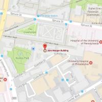
Penn PET Explorer
Perelman School of Medicine at the University of Pennsylvania
John Morgan Building
3620 Hamilton Walk
Philadelphia, PA 19104
Joel Karp, Ph.D.
215-573- 4998
Email Us