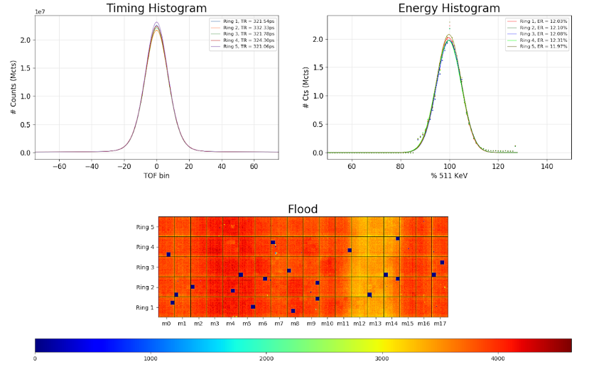
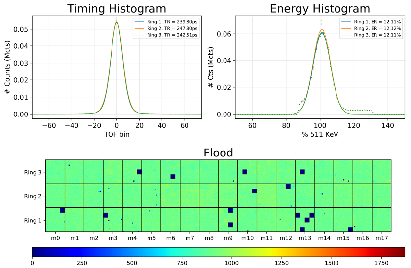
Quality check on the five-ring PennPET Explorer (at room temperature) shows 320 ps timing resolution and 12% energy resolution for all five rings. The flood is uniform for all five rings. Note the slight non-uniformity is due to the geometry of the source.
03/16/2020: Completion of five scanner rings in dedicated imaging suite
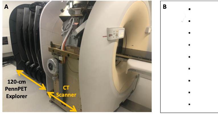
(A) Five rings of the PennPET Explorer built and aligned with a CT in our dedicated imaging suite. (B) Nine point sources, each 12-cm apart, acquired along the length of the five-ring scanner.
06/2019-11/2019: Moving scanner to dedicated imaging suite near clinical space
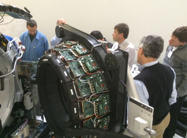
02/2019: Ring 4 undergoing testing with new electronics
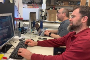
Researchers Mike Parma and Matthew Werner working hard on getting the new scanner ring working
03/2019: Research tracers imaged on the PennPET Explorer
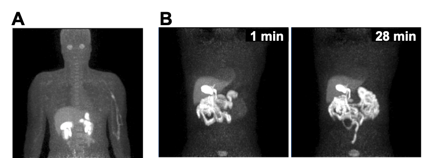
(A) Maximum-intensity projection (3-min scan) of 18F-labeled NOS PET (subject 6). (B) Maximum-intensity projections of 18F-fluortriopride PET (subject 9) for 1-min duration shown at 1 min (left) and 28 min (right) after drinking Ensure (Abbott Laboratories) to stimulate emptying of radiotracer from gallbladder. (Pantel et. al.)
03/2019: Fully Dynamic FDG Human Volunteer Imaged
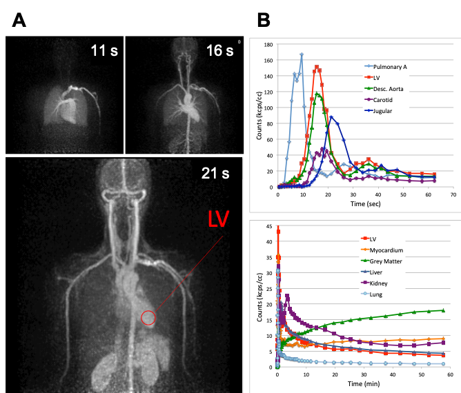
(A) 18F-FDG PET maximum-intensity projections of subject 7, each 1 s in duration, at 3 time points from dynamic scan. (B) Time–activity curves of blood input function measured at several vessels over first minute after injection, and time–activity curves of major organs over first hour after injection. LV = left ventricle. (Pantel et. al.)
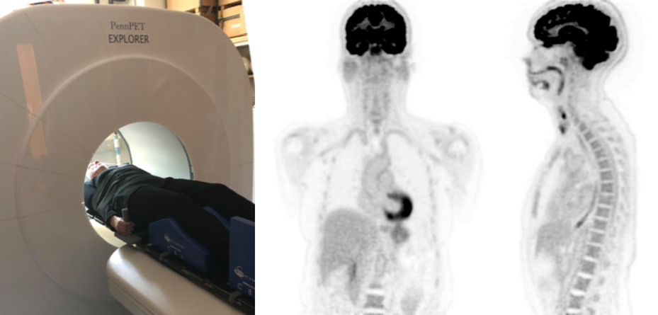
12/2018: First Human Cancer Patient Imaged
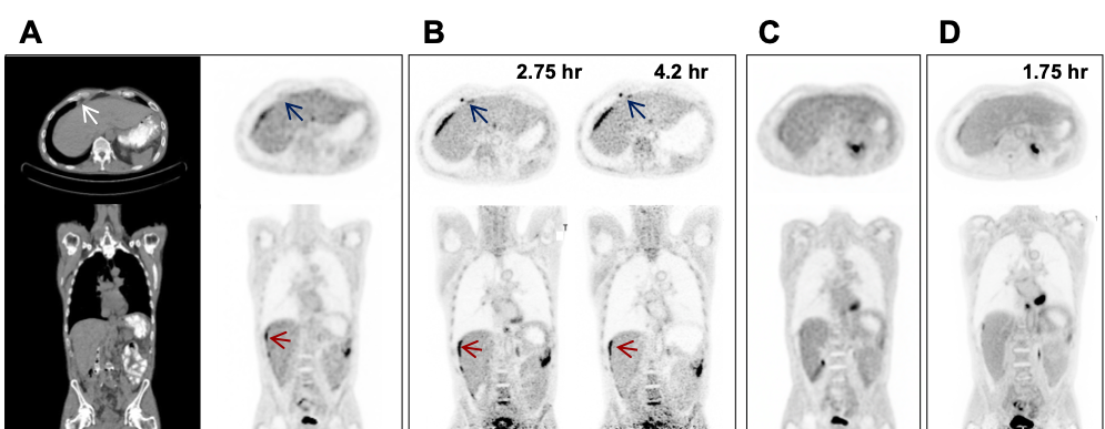
Clinical 18F-FDG PET/CT images (transverse and coronal) from subject 5, with metastatic colon cancer, acquired with standard clinical protocol. (B) PennPET image acquired 2.75 and 4.2 h after injection (10-min scans). Matched coronal and transverse slices are shown. Red arrows denote perihepatic disease; blue arrows denote epiphrenic lymph node. (C) Follow-up clinical scan at 3 mo (subject 10). (D) Corresponding PennPET image (20-min scan) demonstrating improvement in perihepatic disease and epiphrenic lymph node. (Pantel et. al.)
09/2018: Second FDG Human Volunteer on PennPET Explorer at 250 ps
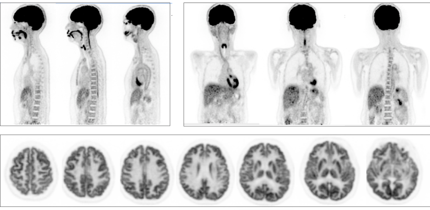
18F-FDG PET images of the second human subject imaged on the PennPET Explorer (10-min scan). Sagittal (top left), and coronal (top right) whole body images (2-mm slice), along with transverse images (bottom) of the patient’s brain positioned in the center of the AFOV. (Karp et al.)
09/2018: Cooled Three Ring Scanner to 5˚C (250 ps)
Quality check on the three-ring PennPET Explorer after cooling the system to 5˚C shows a 250 ps timing resolution and 12% energy resolution for all three rings. The flood is uniform for all three rings.
08/2018: First FDG Human Volunteer Imaged on PennPET Explorer
Image of first human subject being imaged on the PennPET Explorer (left) and resulting coronal (middle) and sagittal (right) reconstructed images. (Karp et al.)
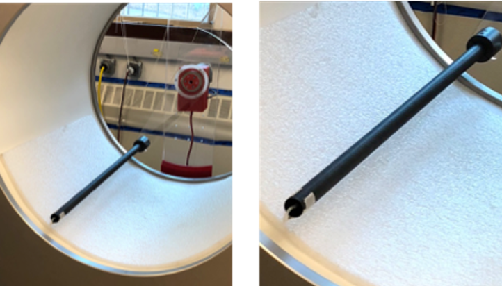
05/2018: First norm taken with a rotating line source
Rotating line source in a carbon fiber tube used to acquire normalization data
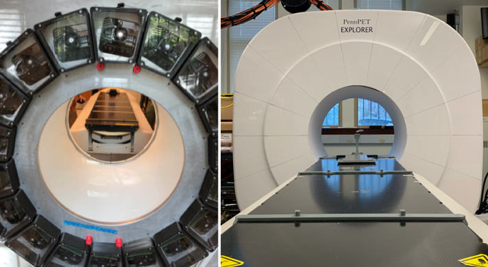
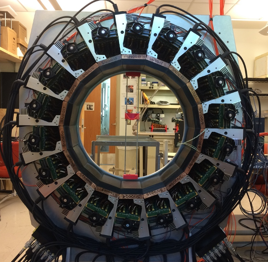
05/2018: Three-Ring PennPET Explorer Built
Three rings of the scanner fully built
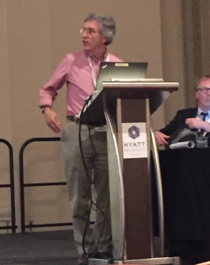
10/2017: Professor Joel Karp at the 2017 IEEE MIC conference
Professor Joel Karp presenting early results from the first two rings of the PennPET Explorer at the 2017 IEEE MIC conference in Atlanta GA.
08/2017: First Ring of PennPET Explorer Built
First ring of scanner fully built. Shown from the back of the scanner.
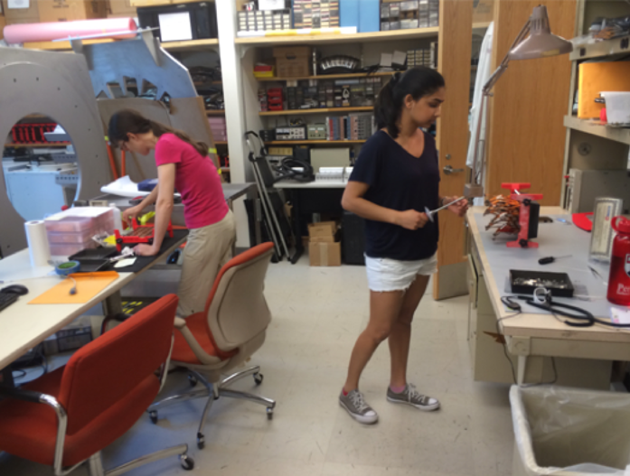
12/2016: Work on building scanner modules continues
Expert technicians, Amy and Varsha, work on building modules of the scanner
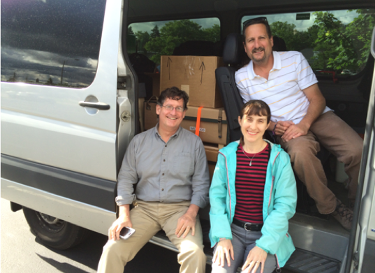
05/2016: The first Philly to Cleveland trip to pick up materials for the scanner
Amy, Michael, and Randy make the first drive to Cleveland to pick up materials for the scanner
02/2016: The formation of PennPET Explorer team
Michael Geagan and Gerd Muehllehner were invited to found the PennPET Explorer team as key members.
11/2015: First visit to the Philips facilities in Cleveland
Joel Karp, Simon Cherry, Ramsey Badawi, and others visit the facility in Cleveland where the Philips Vereos is built.
Contact Us
Penn PET Explorer
Perelman School of Medicine at the University of Pennsylvania
John Morgan Building
3620 Hamilton Walk
Philadelphia, PA 19104