
Ongoing Research
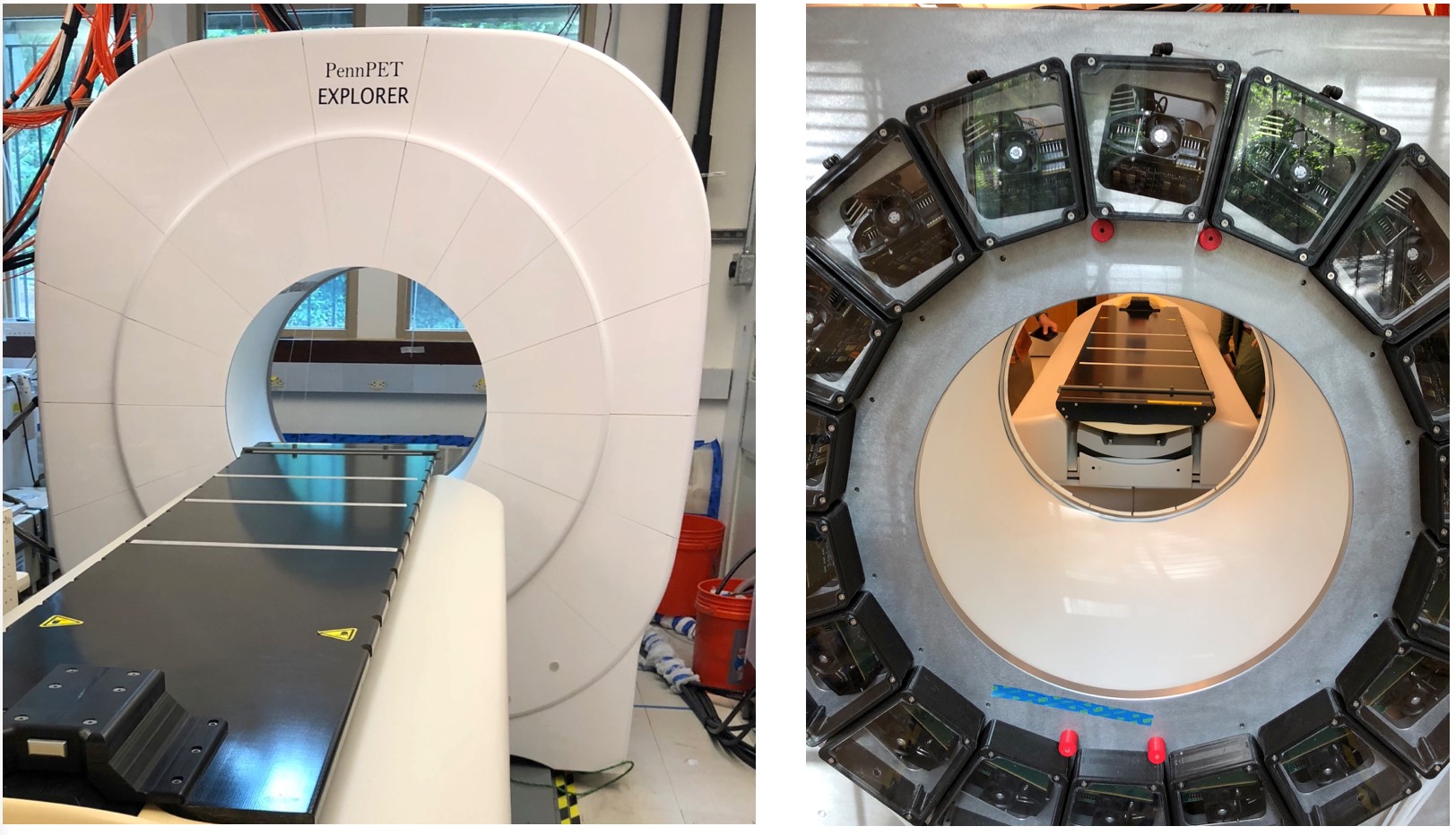
The PennPET Explorer is a whole-body imager that can image the major body organs simultaneously with higher sensitivity than that of commercial devices. The device is scalable in axial length and was initially tested in prototype form with 3 rings and axial field-of-view of 64 cm. (see figure). The technology of the PennPET Explorer is based on the Philips silicon photomultiplier, and the system achieves a time-of-flight (TOF) resolution of 250 ps and a spatial resolution of 4.0 mm (FWHM). (Karp et. al. 2019)
We have recently completed a pilot human imaging study with each study designed to test specific capabilities of the device. (Karp et. al 2019, in press) We demonstrated the ability to scan for shorter duration, or, alternatively with less activity, without a compromise in image quality. In clinical patients, the PennPET Explorer better delineates extent of FDG-avid disease. Delayed images, up to 10 half-lives with FDG, reveal biological insight and support the ability to track biologic processes over time. Dynamic imaging studies capture relatively noise-free input functions for kinetic modeling approaches. Additional studies with experimental research radio-tracers illustrate the benefits from the combination of large axial coverage and high sensitivity. These studies provide proof-of-concept for many proposed applications for the PennPET scanner, which is currently being extended with additional rings for an axial FOV of 140 cm. The PennPET is expected to begin operation in the Radiology PET Center in early 2020.
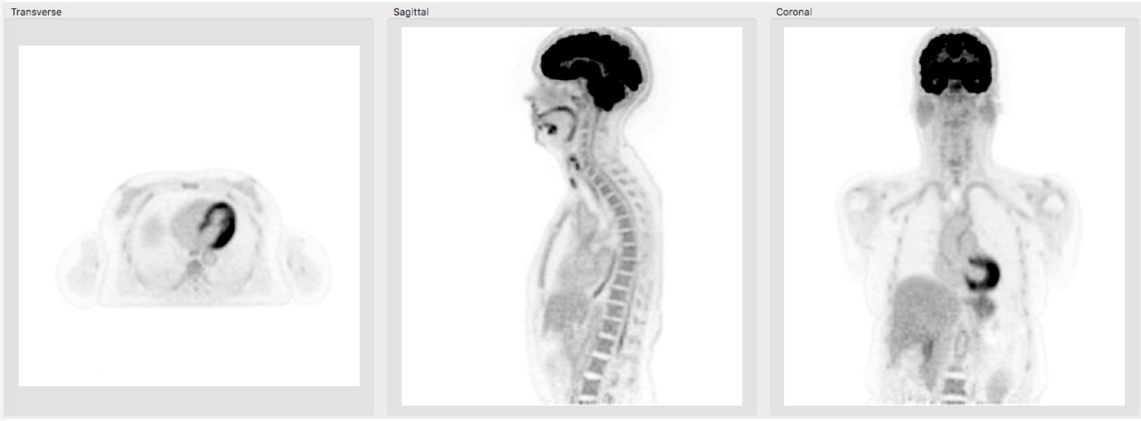
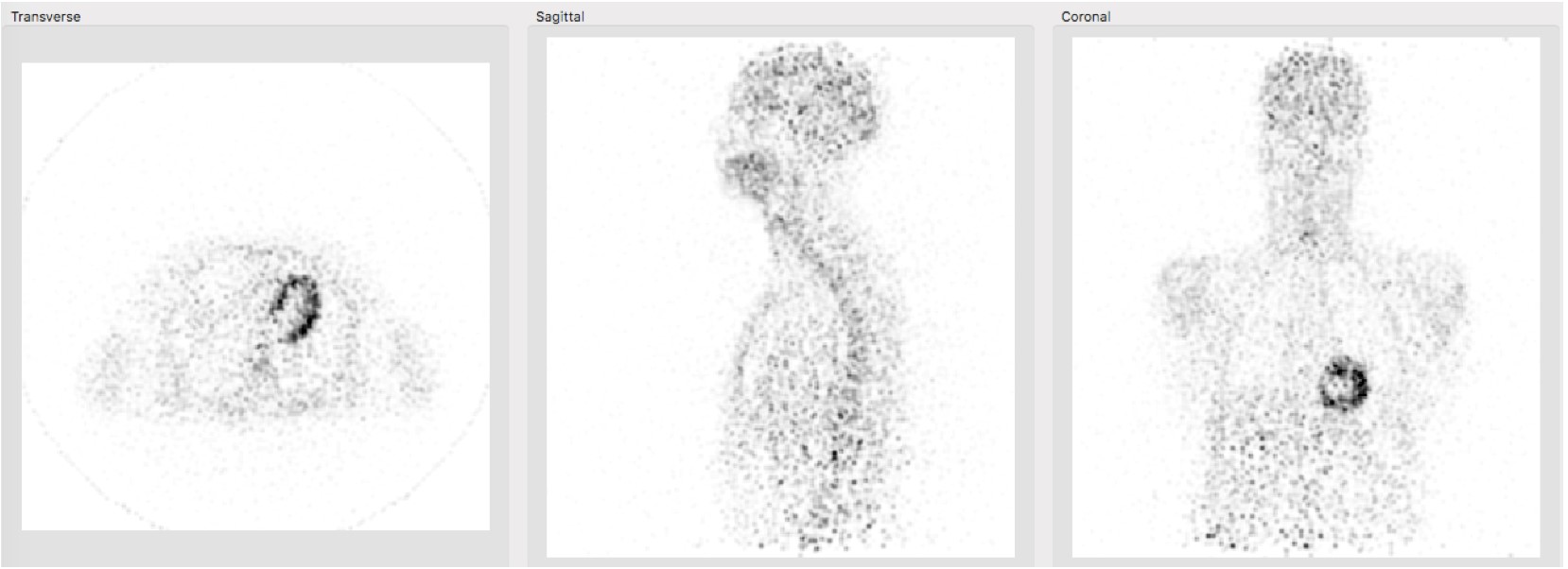
An example of a human study imaged with FDG is shown below. The first scan, acquired at 1.5 hr post-injection. illustrates the excellent image quality – low noise with high structural detail - achieved in a short scan with a single-bed position. The second scan, acquired at 19 hr post-injection illustrates that the very high sensitivity of the PennPET Explorer enables measurement of both fast and slow kinetics, and that valuable information can be extracted from the radiotracer distribution after more than 10 half-lives. This type of study had not been previously possible with commercial PET/CT instruments with standard axial field-of-view.
Selected Publications
- Pantel AR, Viswanath V, Daube-Witherspoon ME, Dubroff JG, Muehllehner G, Parma MJ, Pryma DA, Schubert EK, Mankoff DA, Karp JS. PennPET Explorer: Human imaging on a whole-body imager. J Nucl Med (in press).
- Karp JS, Viswanath V, Geagan MJ, Muehllehner G, Pantel AR, Parma MJ, Perkins AE, Schmall JP, Werner ME, Daube-Witherspoon ME. PennPET Explorer: Design and preliminary performance of a whole-body imager. J Nucl Med September 2019. [Link]
- Viswanath V, Daube-Witherspoon ME, Schmall JP, Surti S, Werner ME, Muehllehner G, Geagan MJ, Perkins AE, Karp JS. Development of PET for total-body imaging. Acta Phys Polonica, vol. 48, pp. 1555-1566, 2017. [Link]
- Simon CR, Jones T, Karp JS, Qi J, Moses WM, Badawi RD. Total-body PET: maximizing sensitivity to create new opportunities for clinical research and patient care. Journal of Nuclear Medicine 59.1 (2018): 3-12.
Contact Us
Penn PET Explorer
Perelman School of Medicine at the University of Pennsylvania
John Morgan Building
3620 Hamilton Walk
Philadelphia, PA 19104
Joel Karp, Ph.D.
215-573- 4998
Email Us