Publications
Numerical observer study of lesion detectability for a long axial field-of-view whole-body PET imager using the PennPET Explorer
Viswanath V, Daube-Witherspoon ME, Karp JS, Surti S.
This work uses lesion detectability to characterize the performance of long axial field of view (AFOV) PET scanners which have increased sensitivity compared to clinical scanners. Studies were performed using the PennPET Explorer, a 70-cm long AFOV scanner built at the University of Pennsylvania, for small lesions distributed in a uniform water-filled cylinder (simulations and measurements), an anthropomorphic torso phantom (measurement), and a human subject (measurement). The lesion localization and detection task was quantified numerically using a generalized scan statistics methodology. Detectability was studied as a function of background activity distribution, scan duration for a single bed position, and axial location of the lesions. For the cylindrical phantom, the areas under the localization receiver operating curve (ALROCs) of lesions placed at various axial locations in the scanner were greater than 0.8 – a value considered to be clinically acceptable (i.e., 80% probability of detecting lesion) – for scan times of 60 s or longer for standard-of-care (SoC) clinical dose levels. 10-mm diameter lesions placed in the anthropomorphic phantom and human subject resulted in ALROCs of 0.8 or greater for scan times longer than 30 s in the lung region and 60 s in the liver region, also for SoC doses. ALROC results from all three activity distributions show similar trends as a function of counts detected per axial location. These results will be used to guide decisions on imaging parameters, such as scan time and patient dose, when imaging patients in a single bed position on long AFOV systems and can also be applied to clinical scanners with consideration of the sensitivity differences.
Close
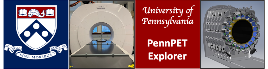
PennPET Explorer: Human imaging on a whole-body imager
Pantel AR, Viswanath V, Daube-Witherspoon ME, Dubroff JG, Muehllehner G, Parma MJ, Pryma DA, Schubert EK, Mankoff DA, Karp JS.
J Nucl Med, vol. 61, pp. 144-151, 2020.
Abstract: The PennPET Explorer, a prototype whole-body imager currently operating with a 64-cm axial field of view, can image the major body organs simultaneously with higher sensitivity than that of commercial devices. We report here the initial human imaging studies on the PennPET Explorer, with each study designed to test specific capabilities of the device. Methods: Healthy subjects were imaged with FDG on the PennPET Explorer. Subsequently, clinical subjects with disease were imaged with 18F-FDG and 68Ga-DOTATATE, and research subjects were imaged with experimental radiotracers. Results: We demonstrated the ability to scan for a shorter duration or, alternatively, with less activity, without a compromise in image quality. Delayed images, up to 10 half-lives with 18F-FDG, revealed biologic insight and supported the ability to track biologic processes over time. In a clinical subject, the PennPET Explorer better delineated the extent of 18F-FDG–avid disease. In a second clinical study with 68Ga-DOTATATE, we demonstrated comparable diagnostic image quality between the PennPET scan and the clinical scan, but with one fifth the activity. Dynamic imaging studies captured relatively noise-free input functions for kinetic modeling approaches. Additional studies with experimental research radiotracers illustrated the benefits from the combination of large axial coverage and high sensitivity. Conclusion: These studies provided a proof of concept for many proposed applications for a PET scanner with a long axial field of view.
Close
PennPET Explorer: Design and preliminary performance of a whole-body imager
Karp JS, Viswanath V, Geagan MJ, Muehllehner G, Pantel AR, Parma MJ, Perkins AE, Schmall JP, Werner ME, Daube-Witherspoon ME.
J Nucl Med, vol. 61, pp. 136-143, 2020.
Abstract: We report on the development of the PennPET Explorer whole-body imager. Methods: The PennPET Explorer is a multiring system designed with a long axial field of view. The imager is scalable and comprises multiple 22.9-cm-long ring segments, each with 18 detector modules based on a commercial digital silicon photomultiplier. A prototype 3-segment imager has been completed and tested with an active 64-cm axial field of view. Results: The instrument design is described, and its physical performance measurements are presented. These include sensitivity of 55 kcps/MBq, spatial resolution of 4.0 mm, energy resolution of 12%, timing resolution of 256 ps, and a noise-equivalent count rate above 1,000 kcps beyond 30 kBq/mL. After an evaluation of lesion torso phantoms to characterize quantitative accuracy, human studies were performed on healthy volunteers. Conclusion: The physical performance measurements validated the system design and led to high-quality human studies.
Close
Total-body PET: Maximizing sensitivity to create new opportunities for clinical research and patient care
Cherry SR, Jones T, Karp JS, Qi J, Moses WW, Badawi RD
Abstract: PET is widely considered the most sensitive technique available for noninvasively studying physiology, metabolism, and molecular pathways in the living human being. However, the utility of PET, being a photon-deficient modality, remains constrained by factors including low signal-to-noise ratio, long imaging times, and concerns about radiation dose. Two developments offer the potential to dramatically increase the effective sensitivity of PET. First by increasing the geometric coverage to encompass the entire body, sensitivity can be increased by a factor of about 40 for total-body imaging or a factor of about 4–5 for imaging a single organ such as the brain or heart. The world’s first total-body PET/CT scanner is currently under construction to demonstrate how this step change in sensitivity affects the way PET is used both in clinical research and in patient care. Second, there is the future prospect of significant improvements in timing resolution that could lead to further effective sensitivity gains. When combined with total-body PET, this could produce overall sensitivity gains of more than 2 orders of magnitude compared with existing state-of-the-art systems. In this article, we discuss the benefits of increasing body coverage, describe our efforts to develop a first-generation total-body PET/CT scanner, discuss selected application areas for total-body PET, and project the impact of further improvements in time-of-flight PET.
Close
Total-body imaging: Transforming the role of positron emission tomography.
Cherry SR, Badawi RD, Karp JS, Moses W, Price P, Jones T.
The first total-body positron emission tomography (TB-PET) scanner represents a radical change for experimental medicine and diagnostic health care.
Close
Development of PET for Total-Body Imaging
Viswanath V, Daube-Witherspoon ME, Schmall JP, Surti S, Werner ME, Muehllehner G, Geagan MJ, Perkins AE; Karp JS
Acta Physica Polonica B . 2017, Vol. 48 Issue 10, p1555-1566. 12p.
Abstract: PET imaging is a key diagnostic tool used clinically to follow and treat disease. While static FDG scans are routine in the clinic, dynamic imaging of disease-specific tracers is important to provide a more precise measure of treatment response. Commercial scanners have limited axial field-of-view and, therefore, we are building a 70 cm long axial FOV TOF PET/CT scanner to enable whole-body dynamic imaging with very high sensitivity. Our scanner is based on detectors with digital SiPMs to provide 300 ps, or better, timing resolution. In this paper, we describe the design and expected performance of this system that will be used for clinical and translational research at the University of Pennsylvania.
Close
Parallax error in long-axial field-of-view PET scanners — a simulation study.
Schmall JP, Karp JS, Werner ME, Surti S.
There is a growing interest in the design and construction of a PET scanner with a very long axial extent. One critical design challenge is the impact of the long axial extent on the scanner spatial resolution properties. In this work, we characterize the effect of parallax error in PET system designs having an axial field-of-view (FOV) of 198 cm (total-body PET scanner) using fully-3D Monte Carlo simulations. Two different scintillation materials were studied: LSO and LaBr3. The crystal size in both cases was 4 × 4 × 20 mm3. Several different depth-of-interaction (DOI) encoding techniques were investigated to characterize the improvement in spatial resolution when using a DOI capable detector. To measure spatial resolution we simulated point sources in a warm background in the center of the imaging FOV, where the effects of axial parallax are largest, and at several positions radially offset from the center. Using a line-of-response based ordered-subset expectation maximization reconstruction algorithm we found that the axial resolution in an LSO scanner degrades from 4.8 mm to 5.7 mm (full width at half max) at the center of the imaging FOV when extending the axial acceptance angle (α) from ±12° (corresponding to an axial FOV of 18 cm) to the maximum of ±67°—a similar result was obtained with LaBr3, in which the axial resolution degraded from 5.3 mm to 6.1 mm. For comparison we also measured the degradation due to radial parallax error in the transverse imaging FOV; the transverse resolution, averaging radial and tangential directions, of an LSO scanner was degraded from 4.9 mm to 7.7 mm, for a measurement at the center of the scanner compared to a measurement with a radial offset of 23 cm. Simulations of a DOI detector design improved the spatial resolution in all dimensions. The axial resolution in the LSO-based scanner, with α = ± 67°, was improved from 5.7 mm to 5.0 mm by incorporating a two-layer DOI detector. These results characterize the maximum axial blurring for a fully open 2 m long PET scanner and demonstrate that large sensitivity gains are possible with a modest reduction in resolution when using current clinical detector technology with no DOI capability.
Close
Impact of detector design on imaging performance of a long axial field-of-view, whole-body PET scanner.
Surti S, Karp JS.
Current generation of commercial time-of-flight (TOF) PET scanners utilize 20–25 mm thick LSO or LYSO crystals and have an axial FOV (AFOV) in the range of 16–22 mm. Longer AFOV scanners would provide increased intrinsic sensitivity and require fewer bed positions for whole-body imaging. Recent simulation work has investigated the sensitivity gains that can be achieved with these long AFOV scanners, and has motivated new areas of investigation such as imaging with a very low dose of injected activity as well as providing whole-body dynamic imaging capability in one bed position. In this simulation work we model a 72 cm long scanner and prioritize the detector design choices in terms of timing resolution, crystal size (spatial resolution), crystal thickness (detector sensitivity), and depth-of-interaction (DOI) measurement capability. The generated list data are reconstructed with a list-mode OSEM algorithm using a Gaussian TOF kernel that depends on the timing resolution and blob basis functions for regularization. We use lesion phantoms and clinically relevant metrics for lesion detectability and contrast measurement. The scan time was fixed at 10 min for imaging a 100 cm long object assuming a 50% overlap between adjacent bed positions. Results show that a 72 cm long scanner can provide a factor of ten reduction in injected activity compared to an identical 18 cm long scanner to get equivalent lesion detectability. While improved timing resolution leads to further gains, using 3 mm (as opposed to 4 mm) wide crystals does not show any significant benefits for lesion detectability. A detector providing 2-level DOI information with equal crystal thickness also does not show significant gains. Finally, a 15 mm thick crystal leads to lower lesion detectability than a 20 mm thick crystal when keeping all other detector parameters (crystal width, timing resolution, and DOI capability) the same. However, improved timing performance with 15 mm thick crystals can provide similar or better performance than that achieved by a detector using 20 mm thick crystals.
Close
Contact Us
Penn PET Explorer
Perelman School of Medicine at the University of Pennsylvania
John Morgan Building
3620 Hamilton Walk
Philadelphia, PA 19104
Joel Karp, Ph.D.
215-573- 4998
Email Us