Magnetic Resonance Imaging (MRI) & Functional Magnetic Resonance Imaging (fMRI)
Magnetic Resonance Imaging (MRI) & Functional Magnetic Resonance Imaging (fMRI)
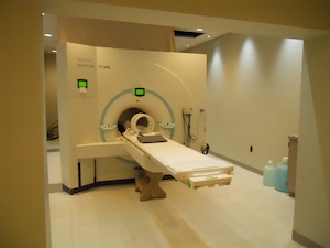
Magnetic resonance imaging (MRI) is a medical imaging technique that is used to form high-resolution pictures regions of the body such, as the brain. MRI scanners employ strong magnetic fields, magnetic field gradients, and radio waves to generate images, and do not involve either X-rays or ionizing radiation. As a result, MRI scanning is deemed to be safe for most people. Researchers in the brainSTIM Center often pair MRI data with ‘neuronavigation’ software in order to precisely guide noninvasive neuromodulation to specific areas of the brain.
 Functional magnetic resonance imaging (fMRI) combines the imaging technology of conventional anatomic MRI scanning with the ability to detect local changes in blood flow and oxygenation in the brain. Because brain activity normally leads to local changes in brain blood flow and oxygenation, measuring these changes while subjects perform behavioral tasks allows investigators to map regions of the brain that engage in specific mental activities. Moreover, it is also possible to map which areas of the brain are simultaneously active even when a person is not performing tasks (also known as the resting state), offering valuable insight into which regions of the brain are functionally connected with one another. Investigators in the brainSTIM Center are also able to apply noninvasive neuromodulation (TMS and tDCS) during fMRI, allowing for manipulation of activity in brain sites and networks which can then be measured using imaging.