Measurement for Rat Glenoid Cartilage Thickness- High Frequency Ultrasound
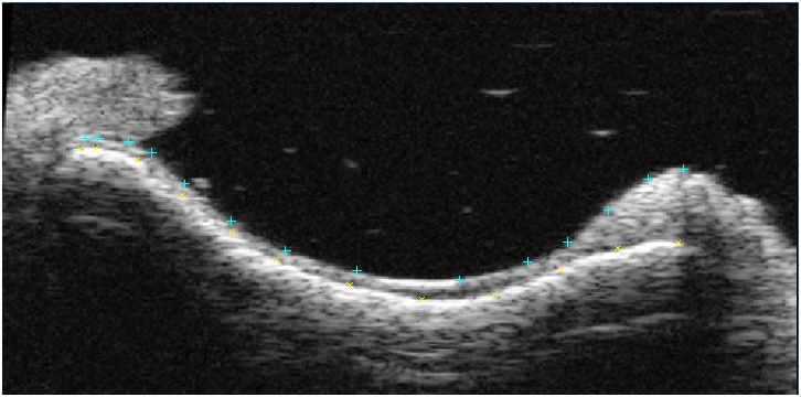
Clinically, complete supraspinatus and infraspinatus rotator cuff tendon tears disrupt the normal balance of forces at the joint, resulting in superior and antero-superior translation of the humeral head and subsequently, altered loading of the glenoid. A correlation between rotator cuff tears and glenohumeral cartilage degeneration has also been observed. Utilizing our rat shoulder model of tendon injury, we plan to study the mechanism by which tendon injuries lead to permanent joint damage, particularly articular cartilage degeneration. Unfortunately, current methods for measuring cartilage thickness, such as needle indentation and histology, are destructive to the tissue and limited in their accuracy and repeatability. Therefore, in conjunction with our collaborators at the University of Pennsylvania's Ultrasound Research Lab, we have developed a non-destructive technique using high frequency, high accuracy ultrasound imaging in order to quantify cartilage thickness in the glenoid of the shoulder (Figure 1 and Figure 2).
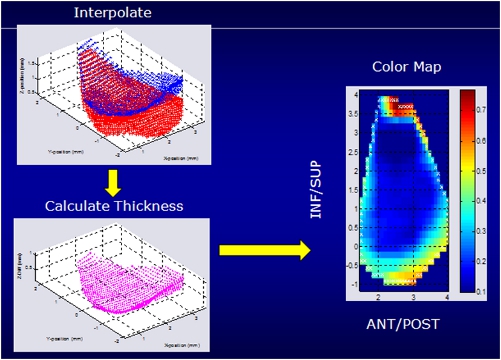
Preliminary results have shown that detachment of the postero-superior dynamic restraints (supraspinatus and infraspinatus) resulted in decreases in glenoid cartilage thickness localized to the inferior regions. Future studies will examine additional time points after rotator cuff injury to examine the progression of cartilage changes as well as tissue mechanical and biological properties, in order to gain insight into the relationships between cuff tears and joint damage.