Ultrasound
Tendons server a primarily mechanical role; consequently, the mechanical properties of tendons are important to their function. The primary load-bearing structure in tendon is collagen, and the organization of the collagen network is known to affect the tendon's mechanical properties. Following injury, the collagen network becomes disorganized, but as the tendon heals over time, the organization of the collagen network improves. Thus, for injured tendon, the level of collagen organization indicates how well the tendon has healed, and therefore, a measure of collagen organization could be used to judge when a tendon has healed enough so that normal activity can be resumed following tendon injury. Unfortunately current methods for measuring tendon organization, such as polarized light analysis, are destructive. There is therefore a need to develop non-destructive and, preferably, non-invasive measures of collagen organization.
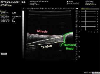
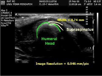
Our collaborators, at the University of Pennsylvania's Ultrasound Research Lab are working with us to develop an ultrasound technique for quantifying collagen organization. To date, we have successfully collected both in-vivo images (LEFT image) and in-vitro (RIGHT image higher resolution) of the rat shoulder seen below.
Currently, we are planning to study changes in tendon organization following collagenase injection.