Last week, researchers and clinicians from around the world, including many from Penn, joined in Philadelphia for the 2024 Alzheimer's Association International Conference (AAIC) to discuss recent advances in Alzheimer's disease and related dementias.
Some highlights of Penn presenters from this year’s conference are featured below:
- Eddie Lee Kicks off Plenary Sessions with “Neuropathology in a Multidisciplinary Age”
-
High-resolution postmortem MRI help paints a picture of co-pathologies in neurodegenerative diseases
-
Short tandem repeats as novel genetic driver of Alzheimer’s disease
-
Comparison of plasma p-tau217 and Aβ42/Aβ40 biomarkers by race to detect Alzheimer’s disease
Eddie Lee Kicks off Plenary Sessions with “Neuropathology in a Multidisciplinary Age”
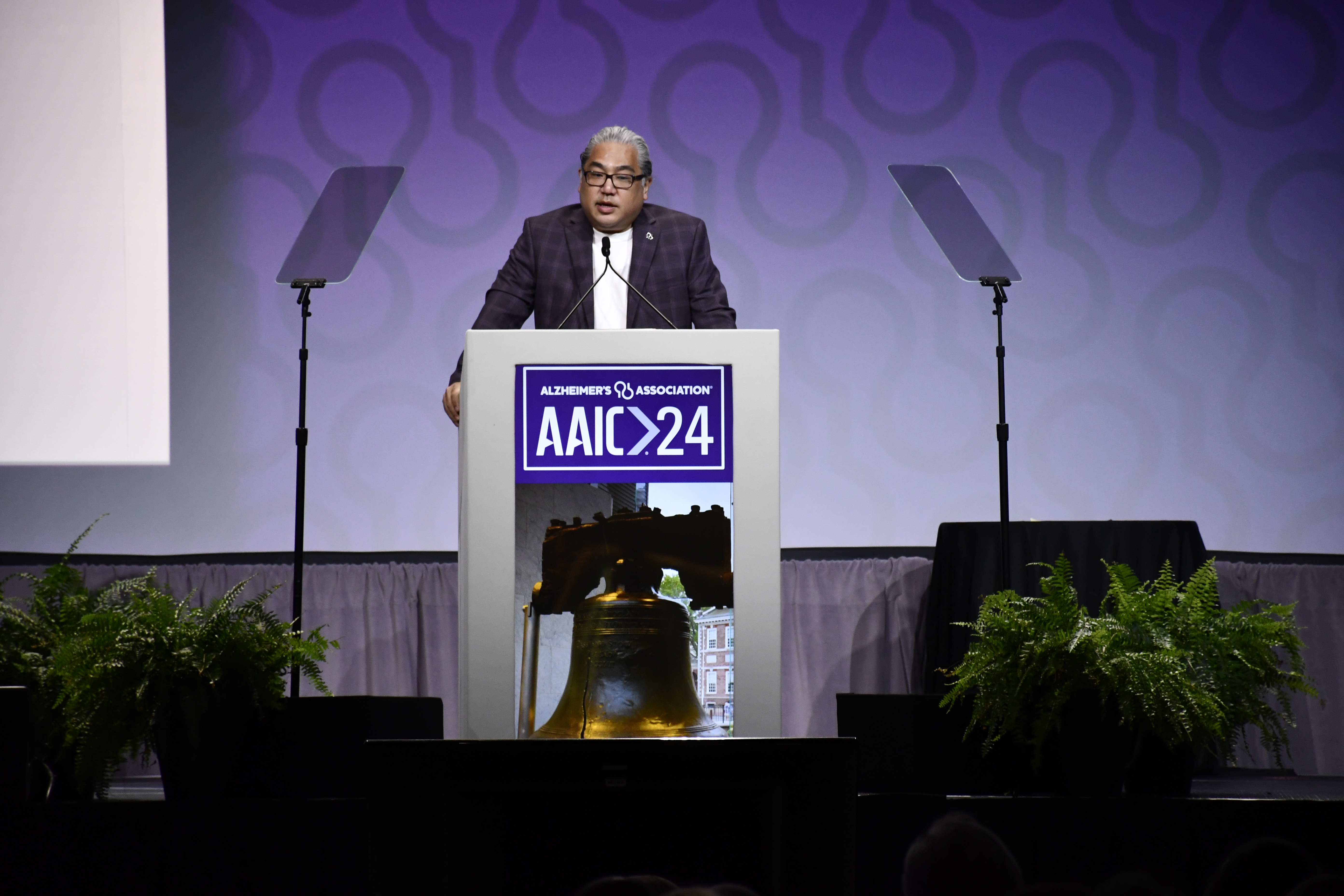 Eddie Lee, MD, PhD, Co-Director of the Institute on Aging, kicked off the plenary sessions at this year’s AAIC with a presentation titled “Neuropathology in a Multidisciplinary Age.” His lecture discussed neuropathology, the study of diseases through brain tissue analysis, and how it intersects with a wide variety of other disciplines to inform our understanding of Alzheimer’s disease and related dementias (ADRD).
Some highlights from his talk included research done here at the University of Pennsylvania including the foundational work of Virginia M.-Y. Lee, PhD and John Q. Trojanowski, MD, PhD, marrying pathology and biochemistry. He also covered pathology and genetics, highlighting how some rare mutation cases such as Valosin Containing Protein (VCP) and Annexin A11 (ANXA11) lead to insights into ADRD. Additional topics included how neuropathology intersects with the environmental sciences and structural biology. This comprehensive talk covering the vast components of the field explained not only how neuropathology serves as the ground truth, but how it can hold the key to many breakthroughs when combined with insights from other disciplines in the field.
"I am honored that the Alzheimer's Association and the Scientific Planning Committee invited me to give this plenary talk at AAIC,” said Dr. Lee. “I hope to represent Philadelphia and highlight the many achievements in ADRD research which were rooted in human neuropathology and amplified by a multidisciplinary approach."
AAIC Plenary Sessions address the field’s most crucial and compelling topics and are delivered by leaders in the field. Eddie Lee, MD, PhD, is an Associate Professor of Pathology and Laboratory Medicine at the Perelman School of Medicine at the University of Pennsylvania and Associate Director of the Penn Alzheimer's Disease Research Center.
High-resolution postmortem MRI help paints a picture of co-pathologies in neurodegenerative diseases
Currently, co-pathologies are not reliably detected with in vivo imaging, making it difficult for clinicians to accurately diagnose the cause of cognitive impairment in individual patients and to identify those most likely to benefit from treatment with emergent drugs that target Alzheimer’s disease pathology. The efficacy of in vivo biomarkers can be enhanced by combined analysis of regional measures of neurodegeneration, such as cortical thickness, derived from brain MRI (ex vivo or in vivo) and pathological measures obtained at autopsy. Such analysis can help identify distinct spatial patterns of disease progression linked to AD-specific pathologies (i.e. β-amyloid plaques and phosphorylated tau protein tangles) and common co-pathologies, however, limited work has been done in this area due to lack of datasets and centers with such imaging resources.
Recently, researchers at Penn Medicine have presented a first-of-its-kind novel dataset comprised of a very large heterogeneous cohort of over 150 human brain specimens spanning Alzheimer's disease and related dementias such as cerebrovascular disease, Limbic-predominant age-related TDP-43 encephalopathy (LATE), and Lewy body disease (LBD) amongst others. The computational pipeline developed by this team helps parcellate the whole-brain hemispheres in native subject-space resolution using a variety of standard brain atlases which enables them to understand the local regional underpinnings of structural morphometry with the underlying histology-derived markers. The study aims to enable scientific advancements in ex vivo imaging and thereby inform better in vivo computational biomarkers with the eventual goal of understanding co-morbidity in ADRD.
"The presented data and computational pipelines opens up exciting avenues for both clinicians and scientists to study structure-pathology associations in postmortem imaging which would then help inform in vivo biomarkers,” said Pulkit Khandelwal (pictured far right), a PhD candidate in the Penn Image Computing and Science (PICSL) at the University of Pennsylvania who presented his work during a Featured Research Session.

“I'm very excited to have had the opportunity to present this work at AAIC as it gives a large platform for scientists and engineers like me who develop computational models to connect, discuss and brainstorm ideas to answer clinical questions and thereby help inform better and precise computational biomarkers," he said, reflecting on his experience.
Short tandem repeats as novel genetic driver of Alzheimer’s disease
While there have been many studies looking at simple mutations in the genome, much of the genetic risk of Alzheimer’s disease remains unknown. With this in mind, research by Penn Medicine physician-scientist Michael Guo, MD, PhD, and his team investigates whether a highly repetitive form of genetic mutation called short tandem repeats (STRs) – a mutation associated with neurological diseases such as Huntington’s disease and frontotemporal dementia (FTD) – may also be associate with Alzheimer’s disease (AD).
Using new genome analysis methods, the team studied these STRs in 3,000 individuals and found that individuals carrying a high burden of STR mutations had a more than 3-fold increase risk of odds of developing AD. This finding suggest that STRs are a form of mutation that greatly increases the risk of AD.
“I’m excited about this work since as a clinician, it is frustrating not to be able to better counsel patients and family members on their risk of developing Alzheimer’s disease,” said Dr. Guo. “I hope that this work will shed new light on the genetics of this disease so that we can better predict risk for patients and family members.”
Insights into LATE from postmortem imaging
Limbic-predominant age-related TDP-43 encephalopathy (LATE) is a common co-pathology in Alzheimer’s disease – perhaps as frequent in occurrence as one in three individuals. Unfortunately, unlike Alzheimer’s disease which has several known biomarkers – i.e. PET scans, cerebrospinal fluid, and more recently, plasma – LATE currently has no known reliable biomarkers to its name. At this time, the only way to confirm for certain that an individual patient with AD had LATE co-pathology is through autopsy. However, it is becoming increasingly important to be able to identify when individuals with AD have LATE co-pathology, especially now that treatments for modifying the course of AD are available. These treatment options may have serious side effects and it is unclear how helpful they are for individuals with both AD and LATE.
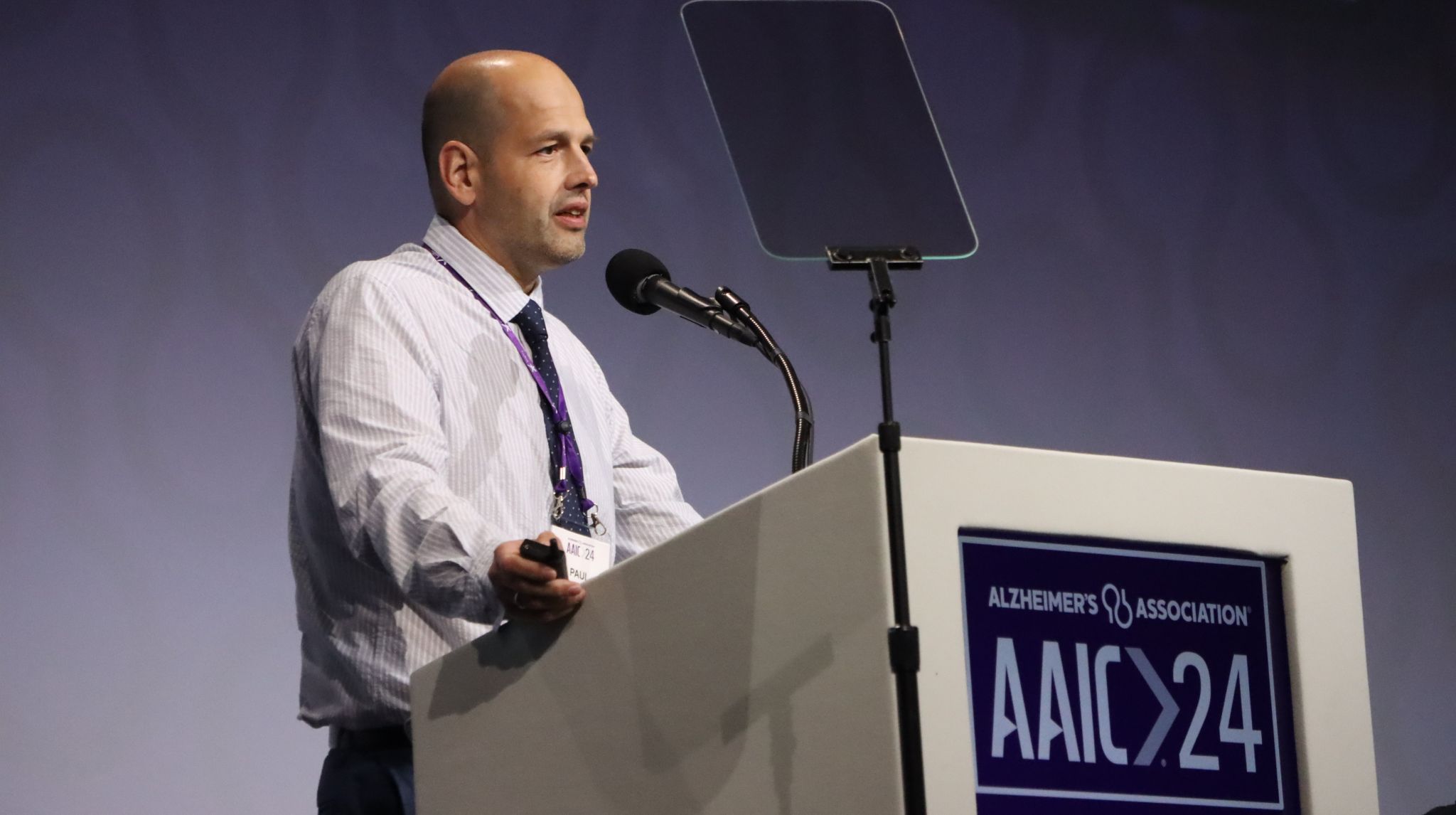 Researchers at the Penn Alzheimer’s Disease Research Center (Penn ADRC), and several other AD research centers, are studying brain scans from brain donors using high-resolution MRI in order to determine whether there are patterns of brain volume loss that differ between AD with and without LATE. Findings suggest that two areas of the brain -- the anterior hippocampus and entorhinal cortex -- are affected more than expected for the level of AD pathology in AD patients with LATE.
During his AAIC Perspectives Session talk, Paul Yushkevich, PhD, a professor of radiology at the University of Pennsylvania, reviewed some of the recent studies on postmortem imaging of LATE and presented novel data from the Penn ADRC that confirms and refines these findings using both postmortem MRI and antemortem MRI scans obtained during life in patients who eventually had autopsy at their center.
“The overall implication of our research and prior studies is that while brain volume loss patterns detected in MRI cannot determine for certain if someone with AD also has LATE, they can be used to provide supporting evidence of LATE co-pathology,” said Dr. Yushkevich.
David Wolk, MD, Co-director of the IOA, co-chaired the panel discussion.
While Alzheimer’s disease (AD) is often associated with issues in memory and thinking, not all patients with the disease have the same experiences or present with the same symptoms.
Researchers at Penn Medicine believe this is partially driven by differences in how tau -- one of the proteins responsible for causing the disease -- accumulates in the brain. “Our research shows how using neuroimaging to identify the structural connections between different parts of each person’s brain can serve as a roadmap to show where tau will accumulate,” explained Christopher Brown, MD, PhD, (pictured far right) a Penn Medicine researcher who presented this work during a Featured Research Session at this year’s AAIC.

This is the first time this type of research has been done at the individual level instead of using a “one-size-fits-all” approach. This method may provide a better way to assess the progression of the disease at the patient-level in both clinical care and clinical trials.
“I'm excited to share this work with the research community at AAIC to promote our new approach and encourage others to implement these models in their studies and clinical trials,” said Dr. Brown.
Comparison of plasma p-tau217 and Aβ42/Aβ40 biomarkers by race to detect Alzheimer’s disease
Blood biomarkers are an excellent tool in detecting Alzheimer’s disease, but before clinical implementation, findings must be validated in diverse populations. A Penn Medicine research study is measuring plasma phosphorylated tau 217 (p-tau217), β-amyloid (Aβ) 1-42/1-40 (Aβ42/Aβ40) ratio, and p-tau217/Aβ42 ratio using Fujirebio Lumipulse – a plasma assay – to test for differences in plasma levels and classification accuracy by self-identified race (White vs. Black), with an established 95% specificity and 95% sensitivity cutpoint for each biomarker.
Findings showed that plasma Aβ42/Aβ40 ratio was higher in individuals identified as Black compared to White, a difference that was confirmed after accounting for variations in Aβ PET burden (standardized uptake value ratio, or SUVR) and area deprivation index. It was also discovered that plasma p-tau217/Aβ42 ratio had the highest accuracy across all groups.
“Blood biomarkers of Alzheimer’s disease offer a non-invasive and cost-effective alternative, with potential to revolutionize clinical care and significantly increase access to diagnosis and treatment,” explained Katheryn Cousins, PhD, a research assistant professor of Neurology at the University of Pennsylvania and author on the study. “However, before these biomarkers can be widely implemented, it is crucial to conduct studies like ours to ensure their accuracy and reliability across diverse populations, including various racial groups.”
The plan is that future research in this area will test biomarkers in a larger sample, and examine the influence of comorbidities on differences in plasma levels and accuracy.
In Alzheimer’s disease (AD), it is not uncommon to have a presence of co-pathologies – a situation that can complicate AD treatment. Previous research from Penn Medicine has shown that Tau (T) – Neurodegeneration (N) mismatch could highlight vulnerabilities and resilience in AD by examining deviations from expected neurodegeneration levels given tau levels. Findings showed that vulnerable phenotypes (N>T) were linked to specific non-Alzheimer’s pathologies (e.g. LATE, vascular disease, etc.). Now, the team’s current research aims to use T-N mismatch to identify distinct spatial-temporal progression patterns of non-AD pathologies – a first-of-its-kind application.
By applying regional T-N mismatch to a machine-learning technique called the Subtype and Stage Inference (SuStaIn), the team identified unique T-N subtypes varying in clinical characteristics and cognitive functions and their progression patterns.
The inferred staging was associated with aging, poorer cognition, and higher white matter hyperintensity -- lesions in the brain that show up as areas of increased brightness when visualized by MRI -- but not AD severity. This suggests that the progression patterns reflect co-pathology trajectories rather than AD progression, potentially benefiting therapeutic strategies in precision medicine.
“My research on T-N mismatch has provided insights into common co-pathologies and their staging in AD, which could potentially benefit clinical trials and therapeutics in the real world,” said Xueying Lyu, a bioengineering PhD student at the University of Pennsylvania working on the study.

“Sharing my findings at AAIC will contribute to a deeper understanding of comorbidities in AD patients. As we move into the era of precision medicine, our approach offers a way to better stratify patients, enhance understanding of co-pathologies and tailor treatments more effectively.”
More from Penn at AAIC:
- A blood test accurately diagnosed Alzheimer’s 90% of the time, study finds – Jason Karlawish, MD, shares his thoughts
- Virginia Lee, PhD leads panel discussion on tau aggregation
- Wildfire smoke linked to increased dementia risk – New researcher presented by Holly Elser, MD, PhD, neurology resident at the University of Pennsylvania
- Plus more!
View Penn Memory Center’s full AAIC 2024 event coverage here.