Translational Research for Precision Rehabilitation
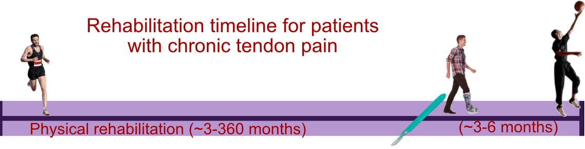
Our research is focused on maximizing patient outcomes following tendon injuries by leveraging precision rehabilitation. Currently, we are investigating how to optimize Achilles tendon loading to promote healing and long-term functional outcomes. This is critical because while orthopaedic surgeons manipulate the tendon loading environment in the operating room, they have very little control over how patients load their injured or healing tendon before and after surgery. In this simple cartoon, we highlight that some patients experience many years of tendon pain before receiving surgical treatment, only to undergo several additional months of physical therapy to promote healing.
By defining precision rehabilitation protocols that promote tendon healing, we expect to 1) reduce the need for surgical treatment in patients with Achilles tendon injuries and 2) when surgery is neccessary, support the healing tendon with therapuetic loading that does not damage the recently repaired tendon. This approach is also impactful because it works within the existing reimbursement environment for physical therapy.
 Achilles tendon ruptures are common and devastating injuries. Although our clinical treatment has improved in recent decades with less than 5% of patients suffering a re-rupture, nearly 2 out of 3 patients suffer at least a 15% permanent deficit in ankle function. Our group's primary focus is 1) understanding what causes these functional deficits and 2) develop new techniques to address the root cause of these functional deficits. Our ongoing research has identified structural changes to the calf muscles that pull on the Achilles tendon as the main reason for these functional deficits. At first it sounds strange that patients who suffer a tendon injury are limited by changes in their muscle. But our studies suggest that the calf muscles rapidly respond to this Achilles tendon injury by remodeling in a biologic attempt to restore the resting tension in the Achilles tendon. We found that during the first 14 weeks following an Achilles tendon rupture, the tendon elongates while the calf muscles remodel in a shorter position. The magnitude of this muscle remodeling appears to explain the amount of functional deficits patients experience — at least early in the rehabilitation process.
Our next set of studies is defining Achilles tendon loading profiles that patients experience throughout their rehabilitation and link these loading profiles with structural and functional outcomes. To accomplish this effort, we have developed innovative measurement techniques to continuously monitor Achilles tendon loading — even when patients are away from the lab and living their lives in the real world. We expect these studies will change how patients rehabilitate their Achilles tendon and calf muscles following injury — bringing a personalized approach to each patient based on their own activity, healing progress, and other factors.
Publications
Baxter JR, Hullfish TJ, Chao W. Functional deficits may be explained by plantarflexor remodeling following Achilles tendon rupture repair: Preliminary findings. J Biomech 2018;79:238–42.
Hullfish TJ, O’Connor KM, Baxter JR. Medial gastrocnemius muscle remodeling correlates with reduced plantarflexor kinetics 14 weeks following Achilles tendon rupture. Journal of Applied Physiology. 2019 Aug 8;127(4):1005–1011.
Hullfish TJ, O’Connor KM, Baxter JR. Gastrocnemius fascicles are shorter and more pennate throughout the first month following acute Achilles tendon rupture. PeerJ. 2019 Apr 23;7:e6788.
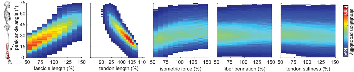
The link between muscle structure and function was defined by the great A.V. Hill in the mid 20th century. However, structure-function relationships are much more complicated in functionally relevant contexts. For example, our lab has found that Achilles tendon ruptures stimulate changes in the shape of the calf muscles. But these structural changes are multi-factorial, which makes it difficult for us to isolate the key structural parameters that truly govern function. To better understand how the structure of our calf muscles and Achilles tendon governs clinically relevant function, we developed a simple musculoskeletal model and simulated these complex interactions on function. In one of our models, we simulated how changes in the Achilles tendon and calf muscles impacts patient function. In these simulations, we found that short muscles and elongated tendons are the most likely causes of functional deficits. Surprisingly, our simulations also found that muscle strength has little to do with the ability to perform a heel raise. This isn't to say that muscle strength isn't important, but rather that strong muscles alone aren't capable of moving your body through movements you enjoy like hiking, jumping, and running. These simulation results highlight the importance of muscle structure on function and support the need to improve treatments that target muscle factors other than muscle strength.
Publications
Baxter JR, Farber DC, Hast MW. Plantarflexor fiber and tendon slack length are strong determinants of simulated single-leg heel raise height. Journal of Biomechanics. 2019 Mar 27;86:27–33.
Baxter JR, Hast MW. Plantarflexor metabolics are sensitive to resting ankle angle and optimal fiber length in computational simulations of gait. Gait & Posture. 2019 Jan 1;67:194–200.
Baxter JR, Hullfish TJ, Chao W. Functional deficits may be explained by plantarflexor remodeling following Achilles tendon rupture repair: Preliminary findings. Journal of Biomechanics. 2018 Oct 5;79:238–242.
 Monitoring patient progress is essential for personalizing rehabilitative care following injuries and surgical treatments. But clinical visits only capture a few instances throughout healing and may miss critical moments that would improve a patient's care. To make this more challenging, COVID-19 has placed additional burdens on monitoring patient throughout rehabilitation. A third important consideration is expanding research access to populations that may not live near large academic centers or aren't able to spend extra time during their clinical visits to participate in research — due to work, childcare, or other obligations. To address this unmet clinical need, our group has developed innovative new technologies and approaches, ranging from wearable sensors to continuously monitor tendon loading to portable strength testing machines.
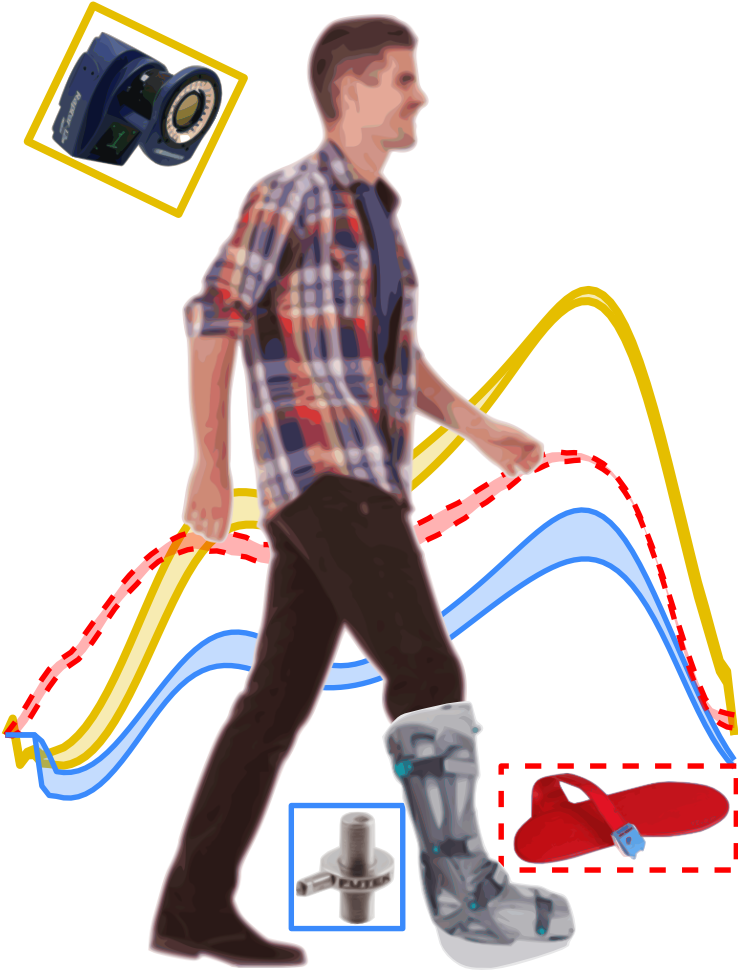
Using an instrumented shoe insole, we have developed a new method to measure Achilles tendon loading while patients walk in an immobilizing boot — those chunky boots you sometimes see people walking around outside. By securing sensors to these boots, we can accurately monitor how much load is being carried by the tendon and how much load is being taken up by the immobilizing boot instead. By using this innovative approach, we will determine the loading profiles that lead to optimal patient outcomes. Additionally, we will also generate new data to improve the design and use of immobilizing boots for patients with Achilles tendon injuries.
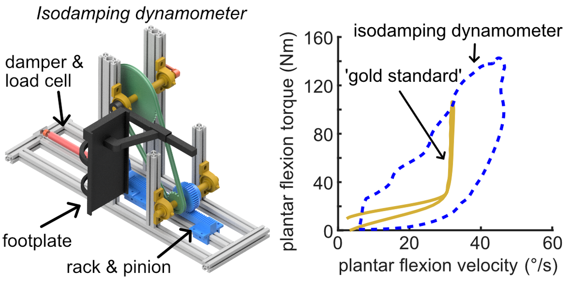 Joint function is typically assessed in the clinic or biomechanics lab using an expensive and heavy device called an isokinetic dynamometer. These dynamometers rely on powerful motors to resist joint work at predefined speeds and are considered to be the gold standard for assessing joint-level function. However, their cost and size make them impractical to utilize in training facilities, smaller clinics, and in the communities where patients and research participants live. To address this current limitation in functional testing, we developed an isodamping dynamometer. Unlike the standard isokinetic dynamometer that uses a powerful motor to resist joint work, our isodamping dynamometer uses inexpensive dampers — similar to what keeps your storm door from slamming when you release it. This design also reduced the weight of the device from around 1,000 pounds to about 40 pounds, which is small and light enough to throw in the back of a car and use at other facilities. We also found that our isodamping dynamometer reliably measures peak joint loads just like the traditional isokinetic dynamometer. But our isodamping dynamometer has improved physiologic relevance because it continuously resists ankle work — not just when it is moving at some pre-defined speed.
Publications
Hullfish TJ, O’Connor KM, Baxter JR. Instrumented immobilizing boot paradigm quantifies reduced Achilles tendon loading during gait. Journal of Biomechanics. 2020 Aug 26;109:109925.
Hullfish TJ, Baxter JR. A simple instrumented insole algorithm to estimate plantar flexion moments. Gait & Posture. 2020 Jun 1;79:92–95.
Drazan JF, Hullfish TJ, Baxter JR. Novel isodamping dynamometer accurately measures plantar flexor function. Journal of Biomechanics. 2020 Oct 9;111:110015.
Funding
Our work is funded by the National Institutions of Health (K01AR075877, R01AR078898, R01AR081062, R21AR081497, P50AR080581) and the American Orthopaedic Foot and Ankle Society.