Biomechanics Core Cartilage Testing
In many cases, the mechanical properties of cartilage change, based on its anatomical location. For example, the thickness and elastic modulus of cartilage within the shoulder joint are not uniform throughout. We can elucidate the properties of the tissue with two assays: 1) high frequency ultrasound; and 2) mechanical testing with a spherical probe.
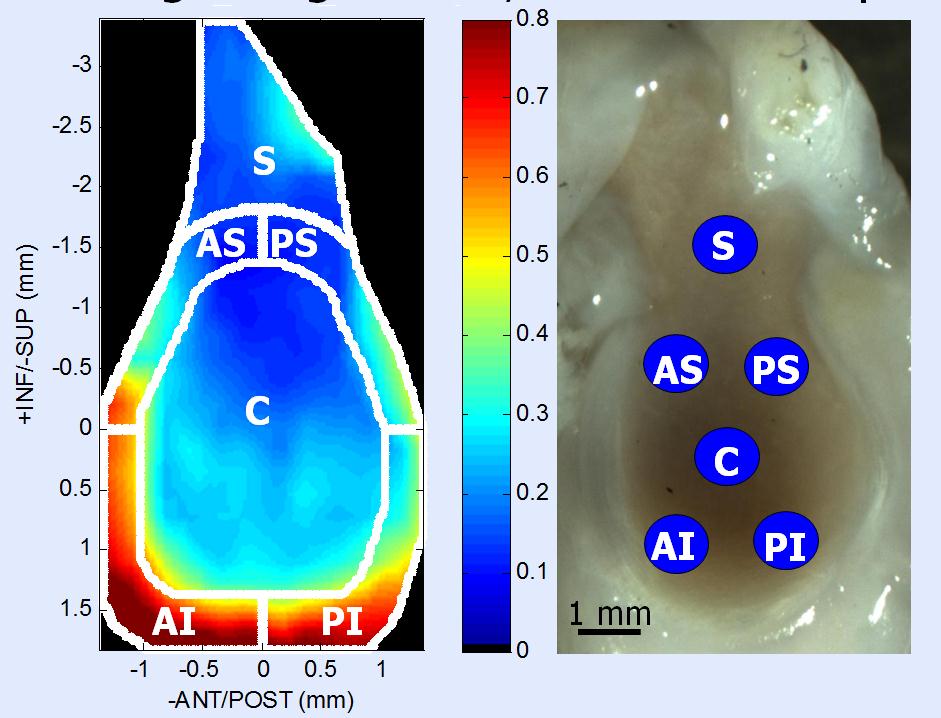
High Frequency Ultrasound: Ultrasound probes used during pregnancy typically have a frequency that ranges between 1.6 -10 MHz. The probes that are used to measure the thickness of cartilage in a small animal must be able to make very fine measurements, and therefore have a much higher frequency, around 55 MHz. In order for measurements to be made, the specimen is submerged into a bath of phosphate buffered saline solution and protease inhibitors, and the specimen is scanned in planes that are 0.25 mm apart. The resulting series of images are then segmented and the thickness of the cartilage can be measured. The images can then be stitched together to create a map of cartilage thickness, as seen in the example below.

Compression Testing: Spherical-tipped probes can be used in conjunction with a uniaxial testing machine to determine the elastic modulus of the cartilage of a specimen. Briefly, the probe can be pressed into various regions of the specimen at a constant rate of displacement. The resulting force-displacement curve can be used to determine the local elasticity of the cartilage. The moduli of the cartilage are subject to change based on factors such as anatomical location and amount of loading the joint experiences on a regular basis. An example of probe placement within a rat glenoid can be seen below.